
Back to article: Microfluidic techniques for separation of bacterial cells via taxis
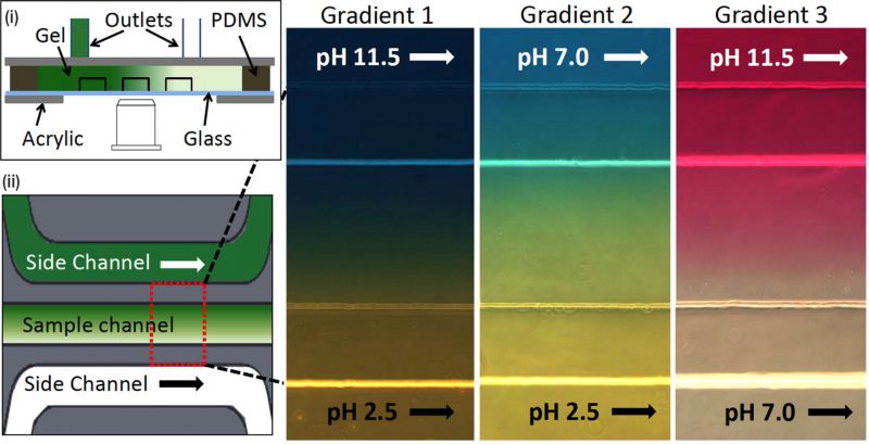
FIGURE 6: Microfluidic device for bacterial pH taxis. (i) Schematic diagram of microfluidic device filled by hydrogel in between three channels and PDMS wall. Acrylic plates clamped the channel attached in the glass slide with the support of PDMS wall. (ii) Schematic diagram of a single sample channel between two side channels. Three different pH gradients (red-dotted rectangular box) were generated by running HCl or NaOH into side channels (green: higher pH, white: lower pH). Reproduced from [48].
48. Zhuang J, Wright Carlsen R, and Sitti M (2015). pH-Taxis of Biohybrid Microsystems. Sci Rep 5: 11403. doi: 10.1038/srep11403