Back to article: Sulforaphane alters the acidification of the yeast vacuole
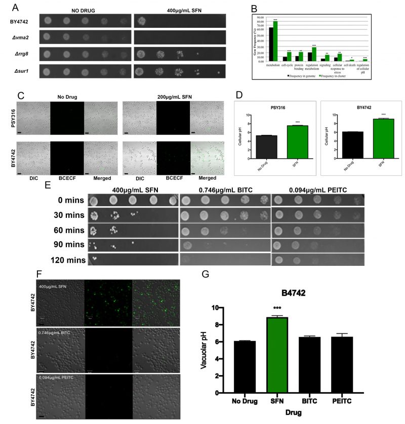
FIGURE 2: SFN alters the acidification of the yeast vacuole.(A) Ten-fold serial dilutions of wild type yeast cells from the BY4742 strain background and of representative mutant strains that were either SFNS or SFNR were plated on synthetic defined (SD) media with 400 µg/mL SFN, and allowed to grow at 30°C for two days. Deletions in genes known to increase vacuolar pH (VMA2) increased the sensitivity of cells to SFN, while deletions in genes known to decrease vacuolar pH (RRG8 and SUR1) increased the resistance of cells to the drug. (B) Functional annotation utilizing gene ontology (GO) terms revealed that our screen had preferentially isolated mutants in genes involved in vacuolar function, especially in vacuolar acidification and/or pH regulation. Asterisks indicate statistical significance of the enrichment of ORFs identified in the screen as compared to their representation in the genome (* p<0.05; ** p<0.01; *** p<0.001). (C) Wild type cells from the PSY316 and the BY4742 strain backgrounds were grown in SD liquid cultures with and without 200 μg/mL of SFN, and were stained with the vacuole specific, pH-sensitive dye, BCECF-AM. Cells grown in SFN were significantly more fluorescent than their counterparts grown in media without drug. (D) The vacuolar pH of the cells imaged in Figure 2C was estimated from a calibration curve that plotted the vacuolar pH of fields of cells grown in APG media titrated to different pH values against the fluorescence intensities measured by the LSM700. Error bars indicate standard deviations for trials with at least three independent cultures. The difference in viabilities was deemed statistically significant by the Student's t-test comparing cells grown in SFN with control cells grown without drug (*** p<0.001). Scale bars indicate a width of 10 µm. (E) 10-fold serial dilutions of wild type yeast cells from the BY4742 strain background cultured in synthetic defined liquid cultures containing the indicated drugs for the indicated time periods (BITC=0.746 µg/mL, PEITC=0.094 µg/mL, SFN=400 µg/mL), were plated on SD media and allowed to grow at 30°C for two days. (F) Wild type cells from the BY4742 strain background were grown in SD liquid cultures containing the indicated drugs for two hours and were stained with the vacuole specific, pH-sensitive dye, BCECF-AM. Cells grown in SFN were fluorescent while their counterparts grown in media with the other drugs were not. Scale bars indicate a width of 10 µm. (G) The vacuolar pH of the cells imaged in Figure 2F was estimated from a calibration curve that plotted the vacuolar pH of cells grown in APG media titrated to different pH values against the fluorescence intensities measured by the LSM700. Error bars indicate standard deviations for trials with at least three independent cultures. The difference in viabilities was deemed statistically significant by the Student's t-test comparing cells grown with the indicated drug with control cells grown without the drug (*** p<0.001).

