Table of contents
Volume 7, Issue 11, pp. 289 - 311, November 2020
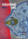
Cover: Colorized electron micrograph showing malaria parasite (right, blue) attaching to a human red blood cell. The inset shows a detail of the attachment point at higher magnification (image by the National Institute of Allergy and Infectious Diseases, National Institutes of Health (USA); the image was modified by MIC). The cover is published under the Creative Commons Attribution (CC BY) license.
Enlarge issue cover
Structural insights into the architecture and assembly of eukaryotic flagella
Narcis-Adrian Petriman and Esben Lorentzen
Reviews |
page 289-299 | 10.15698/mic2020.11.734 | Full text | PDF |
Abstract
Cilia and flagella are slender projections found on most eukaryotic cells including unicellular organisms such as Chlamydomonas, Trypanosoma and Tetrahymena, where they serve motility and signaling functions. The cilium is a large molecular machine consisting of hundreds of different proteins that are trafficked into the organelle to organize a repetitive microtubule-based axoneme. Several recent studies took advantage of improved cryo-EM methodology to unravel the high-resolution structures of ciliary complexes. These include the recently reported purification and structure determination of axonemal doublet microtubules from the green algae Chlamydomonas reinhardtii, which allows for the modeling of more than 30 associated protein factors to provide deep molecular insight into the architecture and repetitive nature of doublet microtubules. In addition, we will review several recent contributions that dissect the structure and function of ciliary trafficking complexes that ferry structural and signaling components between the cell body and the cilium organelle.
Novobiocin inhibits membrane synthesis and vacuole formation of Enterococcus faecalis protoplasts
Rintaro Tsuchikado, Satoshi Kami, Sawako Takahashi and Hiromi Nishida
Research Articles |
page 300-308 | 10.15698/mic2020.11.735 | Full text | PDF |
Abstract
We demonstrate that plasma membrane biosynthesis and vacuole formation require DNA replication in Enterococcus faecalis protoplasts. The replication inhibitor novobiocin inhibited not only DNA replication but also cell enlargement (plasma membrane biosynthesis) and vacuole formation during the enlargement of the E. faecalis protoplasts. After novobiocin treatment prior to vacuole formation, the cell size of E. faecalis protoplasts was limited to 6 μm in diameter and the cells lacked vacuoles. When novobiocin was added after vacuole formation, E. faecalis protoplasts grew with vacuole enlargement; after novobiocin removal, protoplasts were enlarged again. Although cell size distribution of the protoplasts was similar following the 24 h and 48 h novobiocin treatments, after 72 h of novobiocin treatment there was a greater number of smaller sized protoplasts, suggesting that extended novobiocin treatment may inhibit the re-enlargement of E. faecalis protoplasts after novobiocin removal. Our findings demonstrate that novobiocin can control the enlargement of E. faecalis protoplasts due to inhibition of DNA replication.
A novel antibacterial strategy: histone and antimicrobial peptide synergy
Leora Duong, Steven P. Gross and Albert Siryaporn
Microreviews |
page 309-311 | 10.15698/mic2020.11.736 | Full text | PDF |
Abstract
The rate at which antibiotics are discovered and developed has stagnated; meanwhile, antibacterial resistance continually increases and leads to a plethora of untreatable and deadly infections worldwide. Therefore, there is a critical need to develop new antimicrobial strategies to combat this alarming reality. One approach is to understand natural antimicrobial defense mechanisms that higher-level organisms employ in order to kill bacteria, potentially leading to novel antibiotic therapeutic approaches. Mammalian histones have long been reported to have antibiotic activity, with the first observation of their antibacterial properties reported in 1942. However, there have been doubts about whether histones could truly have any such role in the animal, predominantly based on two issues: they are found in the nucleus (so are not in a position to encounter bacteria), and their antibiotic activity in vitro has been relatively weak in physiological conditions. More recent studies have addressed both sets of concerns. Histones are released from cells as part of neutrophil extracellular traps (NETs) and are thus able to encounter extracellular bacteria. Histones are also present intracellularly in the cytoplasm attached to lipid droplets, positioning them to encounter cytosolic bacteria. Our recent work (Doolin et al., 2020, Nat Commun), which is discussed here, shows that histones have synergistic antimicrobial activities when they are paired with antimicrobial peptides (AMPs), which form pores in bacterial membranes and co-localize with histones in NETs. The work demonstrates that histones enhance AMP-mediated pores, impair bacterial membrane recovery, depolarize the bacterial proton gradient, and enter the bacterial cytoplasm, where they restructure the chromosome and inhibit transcription. Here, we examine potential mechanisms that are responsible for these outcomes.


