Table of contents
Volume 7, Issue 12, pp. 312 - 325, December 2020
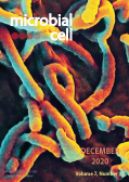
Cover: Scanning electron micrograph of Ebola virus budding from the surface of a Vero cell, an African green monkey kidney epithelial cell line (image by the National Institute of Allergy and Infectious Diseases, National Institutes of Health (USA); the image was modified by MIC). The cover is published under the Creative Commons Attribution (CC BY) license.
Enlarge issue cover
Extracellular vesicles: An emerging platform in gram-positive bacteria
Swagata Bose, Shifu Aggarwal, Durg Vijai Singh and Narottam Acharya
Reviews |
page 312-322 | 10.15698/mic2020.12.737 | Full text | PDF |
Abstract
Extracellular vesicles (EV), also known as membrane vesicles, are produced as an end product of secretion by both pathogenic and non-pathogenic bacteria. Several reports suggest that archaea, gram-negative bacteria, and eukaryotic cells secrete membrane vesicles as a means for cell-free intercellular communication. EVs influence intercellular communication by transferring a myriad of biomolecules including genetic information. Also, EVs have been implicated in many phenomena such as stress response, intercellular competition, lateral gene transfer, and pathogenicity. However, the cellular process of secreting EVs in gram-positive bacteria is less studied. A notion with the thick cell-walled microbes such as gram-positive bacteria is that the EV release is impossible among them. The role of gram-positive EVs in health and diseases is being studied gradually. Being nano-sized, the EVs from gram-positive bacteria carry a diversity of cargo compounds that have a role in bacterial competition, survival, invasion, host immune evasion, and infection. In this review, we summarise the current understanding of the EVs produced by gram-positive bacteria. Also, we discuss the functional aspects of these components while comparing them with gram-negative bacteria.
Maintaining phagosome integrity during fungal infection: do or die?
Mabel Yang, Glenn F.W. Walpole and Johannes Westman
Microreviews |
page 323-325 | 10.15698/mic2020.12.738 | Full text | PDF |
Abstract
Professional phagocytes represent a critical node in innate immunity and tissue homeostasis through their specialized ability to eat, drink, and digest material from the extracellular milieu. The degradative and microbicidal functions of phagocytes rely on the fusion of lysosomes with endosomal compartments such as phagosomes, resulting in the digestion and recycling of internalized prey and debris. Despite these efforts, several particularly dangerous infections result from a class of tenacious pathogens that resist digestion, often surviving and even proliferating within the confines of the phagosomal membrane. One such example, Candida albicans, is a commensal polymorphic fungus that colonizes ~50% of the population and can cause life-threatening infections in immunocompromised patients. Not only can C. albicans survive within phagosomes, but its ingestion by macrophages triggers a yeast-to-hyphal transition promoting rapid intraphagosomal growth (several microns per hour) while imposing a substantial mechanical burden on the phagosomal membrane surrounding the fungus. Preservation of membrane integrity is essential to maintain the hostile internal environment of the phagosome, a functionality of degradative enzymes and oxidative stress. Yet, biological membranes such as phagosomes have a limited capacity to stretch. Using C. albicans as a model intracellular pathogen, our recent work reveals a mechanism by which phagosomes respond to intraphagosomal growth of pathogens by expanding their surface area, and as a result, maintain the integrity of the phagosomal membrane. We hypothesized that this expansion would be facilitated by the delivery and fusion of membrane from extraneous sources with the phagosome. Consistently, macrophages respond to the yeast-to-hyphal transition through a stretch-induced release of phagosomal calcium, leading to recruitment and insertion of lysosomes that accommodate the expansion of the phagolysosome and preserve its integrity. Below, we discuss this calcium-dependent mechanism of lysosome insertion as a means of avoiding phagosomal rupture. Further, we examine the implications of membrane integrity on the delicate balance between the host and pathogen by focusing on fungal stress responses, nutrient acquisition, inflammasome activation, and cell death.


