Table of contents
Volume 8, Issue 4, pp. 73 - 86, April 2021
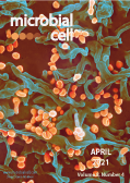
Cover: Scanning electron micrograph of Lassa virus budding off a Vero cell. The Lassa virus causes Lassa hemorrhagic fever in humans and other primates and is endemic in West African countries (image by the National Institute of Allergy and Infectious Diseases, National Institutes of Health (USA); the image was modified by MIC). The cover is published under the Creative Commons Attribution (CC BY) license.
Enlarge issue cover
Neuropathogenesis caused by Trypanosoma brucei, still an enigma to be unveiled
Katherine Figarella
Editorial |
page 73-76 | 10.15698/mic2021.04.745 | Full text | PDF |
Abstract
Trypanosoma brucei is one of the protozoa parasites that can enter the brain and cause injury associated with toxic effects of parasite-derived molecules or with immune responses against infection. Other protozoa parasites with brain tropism include Toxoplasma, Plasmodium, Amoeba, and, eventually, other Trypanosomatids such as T. cruzi and Leishmania. Together, these parasites affect billions of people worldwide and are responsible for more than 500.000 deaths annually. Factors determining brain tropism, mechanisms of invasion as well as processes ongoing inside the brain are not well understood. But, they depend on the parasite involved. The pathogenesis caused by T. brucei initiates locally in the area of parasite inoculation, soon trypanosomes rich the blood, and the disease enters in the so-called early stage. The pathomechanisms in this phase have been described, even molecules used to combat the disease are effective during this period. Later, the disease evolves towards a late-stage, characterized by the presence of parasites in the central nervous system (CNS), the so-called meningo-encephalitic stage. This phase of the disease has not been sufficiently examined and remains a matter of investigation. Here, I stress the importance of delve into the study of the neuropathogenesis caused by
T. brucei, which will enable the identification of pathways that may be targeted to overcome parasites that reached the CNS. Finally, I highlight the impact that the application of tools developed in the last years in the field of neuroscience will have on the study of neglected tropical diseases.
Aeration mitigates endoplasmic reticulum stress in Saccharomyces cerevisiae even without mitochondrial respiration
Huong Thi Phuong, Yuki Ishiwata-Kimata, Yuki Nishi, Norie Oguchi, Hiroshi Takagi and Yukio Kimata
Research Articles |
page 77-86 | 10.15698/mic2021.04.746 | Full text | PDF |
Abstract
Saccharomyces cerevisiae is a facultative anaerobic organism that grows well under both aerobic and hypoxic conditions in media containing abundant fermentable nutrients such as glucose. In order to deeply understand the physiological dependence of S. cerevisiae on aeration, we checked endoplasmic reticulum (ER)-stress status by monitoring the splicing of HAC1 mRNA, which is promoted by the ER stress-sensor protein, Ire1. HAC1-mRNA splicing that was caused by conventional ER-stressing agents, including low concentrations of dithiothreitol (DTT), was more potent in hypoxic cultures than in aerated cultures. Moreover, growth retardation was observed by adding low-dose DTT into hypoxic cultures of ire1∆ cells. Unexpectedly, aeration mitigated ER stress and DTT-induced impairment of ER oxidative protein folding even when mitochondrial respiration was halted by the ro mutation. An ER-located protein Ero1 is known to directly consume molecular oxygen to initiate the ER protein oxidation cascade, which promotes oxidative protein folding of ER client proteins. Our further study using ero1-mutant strains suggested that, in addition to mitochondrial respiration, this Ero1-medaited reaction contributes to mitigation of ER stress by molecular oxygen. Taken together, here we demonstrate a scenario in which aeration acts beneficially on S. cerevisiae cells even under fermentative conditions.


