
Back to article: GFP fusions of Sec-routed extracellular proteins in Staphylococcus aureus reveal surface-associated coagulase in biofilms
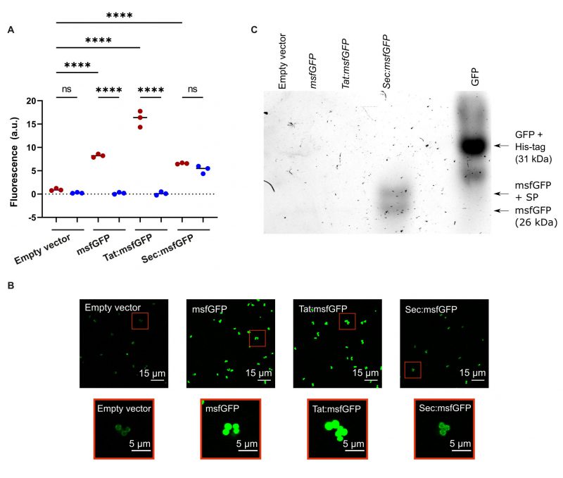
FIGURE 2. (A) Fluorescence intensity from excitation of msfGFP in cell cultures (red circles) or cell-free supernatants (blue circles) of S. aureus expressing msfGFP from the pRMC2 vector. msfGFP was fused to either Tat or Sec signal peptides, no signal peptide, or not expressed at all (empty vector). Black bars indicate group medians. Samples were compared using a one-way ANOVA followed by a Tukey's test; **** denotes a p < 0.0001 significance level and ns denotes no significance. (B) CLSM images of S. aureus cells expressing msfGFP fusions. Red boxes indicate zoomed in images. All fluorescence images had their brightness increased equally using Fiji ImageJ for clear visualisation. (C) In-gel fluorescence of GFP/msfGFP in a native PAGE gel containing supernatants from cultures expressing the empty pRMC2 vector, msfGFP without a signal peptide, or fused to either a Sec or Tat signal peptide.