
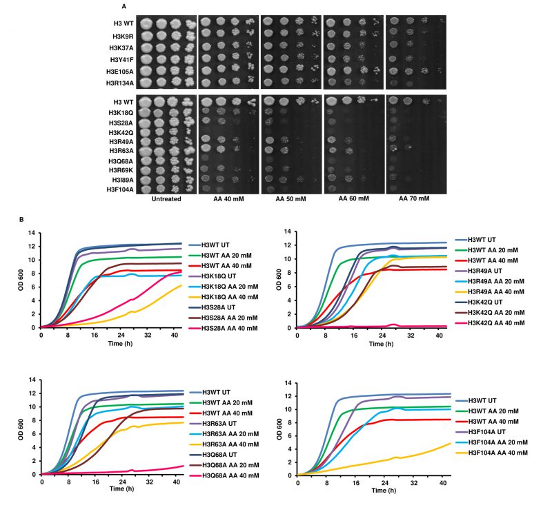
FIGURE 1: Revalidation of the previously identified acetic acid-sensitive histone H3 point mutants. (A) Spot test assay of the wild-type (H3 WT) and the mutants (at 10-fold dilution from left to right) in different concentrations of acetic acid (40, 50, 60 and 70 mM). The untreated plate represents YPD pH 4.5 without any acetic acid and shows the growth of cells at their full potential. Cells were allowed to grow at 30°C, and plates were scanned at 72 h. (B) Growth of the H3 WT and mutant cells in liquid culture under untreated (UT) or acetic acid treated (AA) (20, 40 mM) conditions. The graphs reflect the absorbance values at 600 nm at every 30' for the hours indicated in the x-axis.