
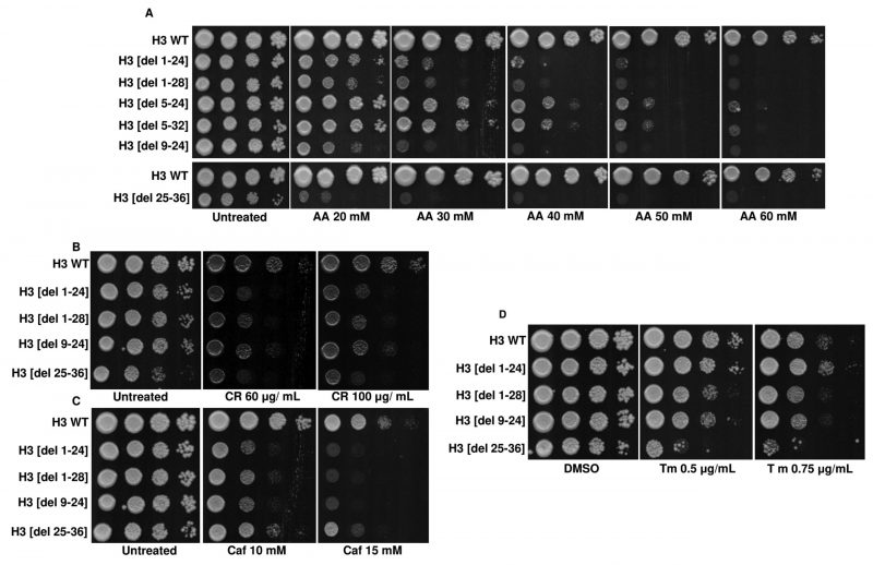
FIGURE 9: Screening of H3 N-terminal deletion mutants identifies acetic acid-hypersensitive mutants. (A) Spot-test assays of the wild-type (H3 WT) and N-terminal tail truncation mutants in different acetic acid concentrations (20, 30, 40, 50, 60 mM). Plates were incubated at 30°C for 72h and photographed. (B) Spot-test assays of the H3 WT and N-terminal tail truncation mutants in different concentrations of Congo red (CR) (60, 100 μg/mL) or (C) Caffeine (Caf) (10, 15 mM). Plates were incubated at 30 °C for 48h (CR) or 72h (Caf) and scanned. (D) Spot-test assays of the H3 WT and N-terminal tail truncation mutants in different concentrations of tunicamycin (Tm) (0.5 μg/mL, 0.75 μg/mL). Images were scanned after 72h.