
Back to article: Expansion of metabolically labelled endocytic organelles and cytoskeletal cell structures in Giardia lamblia using optimised U- ExM protocols
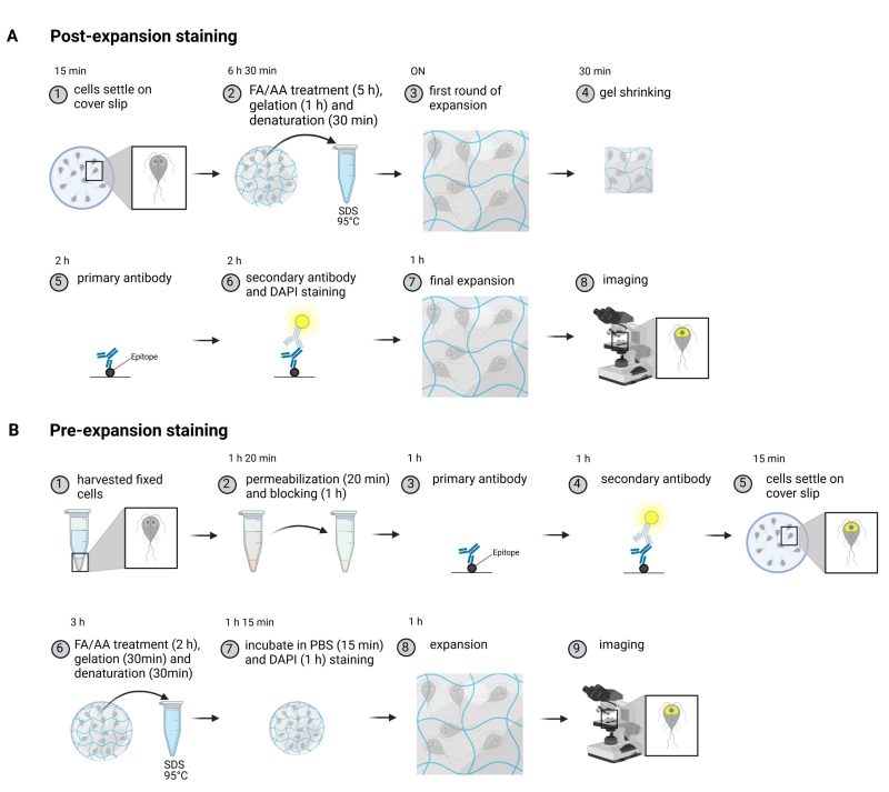
Protocol of a post-and a substantially shortenedpre-expansion staining method. (A) An overview of the post-expansion staining protocol. Briefly, trophozoites of G. lamblia are left to settle on a cover slip (1). After addition of formaldehyde (FA) and acrylamide (AA), cells are embedded into a polymer and proteins are denatured (2). After a first round of expansion (3), the gels are shrunk in PBS (4). Compared to regular fluorescent assays, antibody incubations are longer for both the primary (5) and secondary antibodies requiring up to three hours per incubation (6). During secondary antibody incubation, DNA is stained with DAPI (6). Following the final expansion (7) gels are imaged (8). (B) Cells are harvested and fixed (1) prior to permeabilization and blocking (2). Epitopes are stained with primary (3) and secondary (4) antibodies before settling cells on a cover slip. Stained cells are then anchored during FA and AA treatment. Subsequently, gelation and denaturation are performed (6). After briefly washing the cells in PBS, the DNA is stained with DAPI (7) and after a final expansion step (8), the sample is imaged (9). Created with BioRender.com.