
Back to article: Expansion of metabolically labelled endocytic organelles and cytoskeletal cell structures in Giardia lamblia using optimised U- ExM protocols
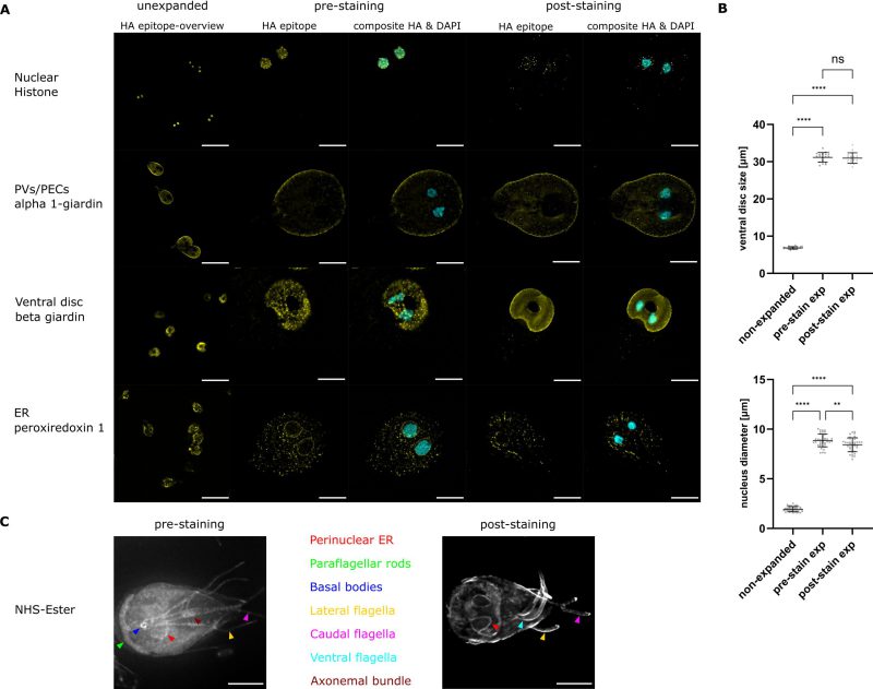
Comparison of a post- and a substantially shortened pre-expansion staining method used to visualise different cell compartments in G. lamblia. (A) Representative widefield light microscopy images of antibody labelled HA epitope tagged proteins expressed in G. lamblia trophozoites. Displayed are overview images of unexpanded cells in the first column. These cells were split off from the samples that were then further treated in the pre-expansion staining ExM method displayed in this figure. Representative cells of the pre- and post-expansion staining methods are depicted in columns 2-5 with the composite images including DNA stain in cyan. On the left the expected localisation and below the tagged protein is indicated. Signal was enhanced for better visibility. Scale bars: 20µm. (B) Expansion factors measured for unexpanded, pre- and post-expansion stained nuclei and ventral discs (n=20 cells). (C) Maximal Z-projection of widefield images of pre- and post-expanded cells treated with NHS-Ester. Different compartments are colour coded. Scale bars: 20 µm.