
Back to article: Expansion of metabolically labelled endocytic organelles and cytoskeletal cell structures in Giardia lamblia using optimised U- ExM protocols
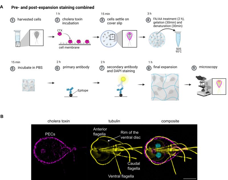
Cholera toxin-dependent metabolic labelling of peripheral endocytic compartments inExM. The workflow of metabolic cholera toxin labelling with subsequent antibody staining is depicted in (A). Briefly, harvested cells (1) are incubated with cholera toxin (CTX) which binds to glycans on cell membranes (2). Cells are allowed to settle on a cover slip (3) before performing the anchoring with formaldehyde (FA) and acrylamide (AA). After gelation and denaturation (4), the gels are briefly washed in PBS (5) before performing tubulin staining with primary (6) and secondary antibodies together with DAPI (7). An example image showing the CTX staining in magenta, tubulin in yellow and a composite including DAPI staining is depicted in part (B). Signal was enhanced for better visibility and colours are displayed in cmyk rather than original fluorophore colour. Scale bar 5 µm. Created with BioRender.com.