Table of contents
Volume 1, Issue 7, pp. 210 - 255, July 2014
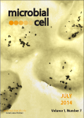
Cover: Sub-inhibitory concentrations of LL-37 enter the bacterial cytosol and interact with DNA. Visualization of LL-37 localization in P. aeruginosa cells using transmission electron microscopy. Bacteria were treated with sub-inhibitory LL-37 and cells were labeled with anti-LL-37 antibody conjugated to Protein G colloidal gold (20 nm) and anti-dsDNA antibody conjugated to Protein G colloidal gold (10 nm). Picture by Dominique H. Limoli and Daniel J. Wozniak (Ohio State University, USA). The cover is published under the Creative Commons Attribution (CC BY) license.
Enlarge issue cover
Sphingolipids and mitochondrial function, lessons learned from yeast
Pieter Spincemaille, Bruno P.A. Cammue, Karin Thevissen
Reviews |
page 210-224 | 10.15698/mic2014.07.156 | Full text | PDF |
Abstract
Mitochondrial dysfunction is a hallmark of several neurodegenerative diseases such as Alzheimer’s disease and Parkinson’s disease, but also of cancer, diabetes and rare diseases such as Wilson’s disease (WD) and Niemann Pick type C1 (NPC). Mitochondrial dysfunction underlying human pathologies has often been associated with an aberrant cellular sphingolipid metabolism. Sphingolipids (SLs) are important membrane constituents that also act as signaling molecules. The yeast Saccharomyces cerevisiae has been pivotal in unraveling mammalian SL metabolism, mainly due to the high degree of conservation of SL metabolic pathways. In this review we will first provide a brief overview of the major differences in SL metabolism between yeast and mammalian cells and the use of SL biosynthetic inhibitors to elucidate the contribution of specific parts of the SL metabolic pathway in response to for instance stress. Next, we will discuss recent findings in yeast SL research concerning a crucial signaling role for SLs in orchestrating mitochondrial function, and translate these findings to relevant disease settings such as WD and NPC. In summary, recent research shows that S. cerevisiae is an invaluable model to investigate SLs as signaling molecules in modulating mitochondrial function, but can also be used as a tool to further enhance our current knowledge on SLs and mitochondria in mammalian cells.
Effect of paraquat-induced oxidative stress on gene expression and aging of the filamentous ascomycete Podospora anserina
Matthias Wiemer, Heinz D. Osiewacz
Research Articles |
page 225-240 | 10.15698/mic2014.07.155 | Full text | PDF |
Abstract
Aging of biological systems is influenced by various factors, conditions and processes. Among others, processes allowing organisms to deal with various types of stress are of key importance. In particular, oxidative stress as the result of the generation of reactive oxygen species (ROS) at the mitochondrial respiratory chain and the accumulation of ROS-induced molecular damage has been strongly linked to aging. Here we view the impact of ROS from a different angle: their role in the control of gene expression. We report a genome-wide transcriptome analysis of the fungal aging model Podospora anserina grown on medium containing paraquat (PQ). This treatment leads to an increased cellular generation and release of H2O2, a reduced growth rate, and a decrease in lifespan. The combined challenge by PQ and copper has a synergistic negative effect on growth and lifespan. The data from the transcriptome analysis of the wild type cultivated under PQ-stress and their comparison to those of a longitudinal aging study as well as of a copper-uptake longevity mutant of P. anserina revealed that PQ-stress leads to the up-regulation of transcripts coding for components involved in mitochondrial remodeling. PQ also affects the expression of copper-regulated genes suggesting an increase of cytoplasmic copper levels as it has been demonstrated earlier to occur during aging of P. anserina and during senescence of human fibroblasts. This effect may result from the induction of the mitochondrial permeability transition pore via PQ-induced ROS, leading to programmed cell death as part of an evolutionary conserved mechanism involved in biological aging and lifespan control.
Exogenous addition of histidine reduces copper availability in the yeast Saccharomyces cerevisiae
Daisuke Watanabe, Rie Kikushima, Miho Aitoku, Akira Nishimura, Iwao Ohtsu, Ryo Nasuno, Hiroshi Takagi
Research Reports |
page 241-246 | 10.15698/mic2014.07.154 | Full text | PDF |
Abstract
The basic amino acid histidine inhibited yeast cell growth more severely than lysine and arginine. Overexpression of CTR1, which encodes a high-affinity copper transporter on the plasma membrane, or addition of copper to the medium alleviated this cytotoxicity. However, the intracellular level of copper ions was not decreased in the presence of excess histidine. These results indicate that histidine cytotoxicity is associated with low copper availability inside cells, not with impaired copper uptake. Furthermore, histidine did not affect cell growth under limited respiration conditions, suggesting that histidine cytotoxicity is involved in deficiency of mitochondrial copper.
A non-proteolytic function of ubiquitin in transcription repression
Ada Ndoja, Tingting Yao
Microreviews |
page 253-255 | 10.15698/mic2014.07.159 | Full text | PDF |
Abstract
Regulation of transcription is vitally important for maintaining normal cellular homeostasis and is also the basis for cellular differentiation, morphogenesis and the adaptability of any organism. Transcription activators, which orchestrate time and locus-specific assembly of complex transcription machinery, act as key players in these processes. One way in which these activators are controlled is by the covalent attachment of the conserved protein, ubiquitin (Ub), which can serve as either a proteolytic or non-proteolytic signal. For a subset of the activators, polyubiquitination-dependent degradation of the activator controls its abundance. In these cases transcription activation can require protein synthesis as well as internal or external stimulus. In contrast, other activators have been reported to undergo mono- or oligoubiquitination that does not lead to protein degradation. The mechanisms by which monoubiquitination of transcription activators affect their activities have been poorly understood. In a recent study, we demonstrated that monoubiquitination of some transcription activators can inhibit transcription by recruiting the AAA+ ATPase Cdc48 (also known in metazoan organisms as p97 or valosin-contain protein, VCP), which then extracts the ubiquitinated activator from DNA.
Mutagenesis by host antimicrobial peptides: insights into microbial evolution during chronic infections
Dominique H. Limoli, Daniel J. Wozniak
Microreviews |
page 247-249 | 10.15698/mic2014.07.157 | Full text | PDF |
Abstract
Antimicrobial peptides (AMPs) are produced by the mammalian immune system to fight invading pathogens. The best understood function of AMPs is to integrate into the membranes of microbes, thereby disrupting and killing cells. However, a recent study [PLoS Pathogens (2014) 10, e1004083] provides evidence that at subinhibitory levels, AMPs promote mutations in bacterial DNA, which enhance bacterial survival. In particular, in the bacterium Pseudomonas aeruginosa, one AMP called LL-37 can promote mutations, which enable the bacteria to overproduce a protective sugar coating, a process called mucoid conversion. P. aeruginosa mucoid conversion is a major risk factor for those suffering from cystic fibrosis (CF), one of the most common lethal, heritable diseases in the US. LL-37 was found to produce mutations by penetrating the bacterial cell and binding to bacterial DNA. It was proposed that LL-37 binding DNA disrupts normal DNA replication and potentiates mutations. Importantly, LL-37 induced mutagenesis was also found to promote resistance to rifampicin in both P. aeruginosa and E. coli. This suggests that AMP-induced mutagenesis may be important for a broad range of chronic diseases and pathogens.
Where antibiotic resistance mutations meet quorum-sensing
Rok Krašovec, Roman V. Belavkin, John A.D. Aston, Alastair Channon, Elizabeth Aston, Bharat M. Rash, Manikandan Kadirvel, Sarah Forbes, Christopher G. Knight
Microreviews |
page 250-252 | 10.15698/mic2014.07.158 | Full text | PDF |
Abstract
We do not need to rehearse the grim story of the global rise of antibiotic resistant microbes. But what if it were possible to control the rate with which antibiotic resistance evolves by de novo mutation? It seems that some bacteria may already do exactly that: they modify the rate at which they mutate to antibiotic resistance dependent on their biological environment. In our recent study [Krašovec, et al. Nat. Commun. (2014), 5, 3742] we find that this modification depends on the density of the bacterial population and cell-cell interactions (rather than, for instance, the level of stress). Specifically, the wild-type strains of Escherichia coli we used will, in minimal glucose media, modify their rate of mutation to rifampicin resistance according to the density of wild-type cells. Intriguingly, the higher the density, the lower the mutation rate (Figure 1). Why this novel density-dependent ‘mutation rate plasticity’ (DD-MRP) occurs is a question at several levels. Answers are currently fragmentary, but involve the quorum-sensing gene luxS and its role in the activated methyl cycle.


