Table of contents
Volume 2, Issue 8, pp. 259 - 304, August 2015
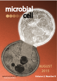
Cover: The cover represents a transmission electron microscopy image of a mutant (pds5-1) yeast cell, showing the nucleus unable to divide at non-permissive temperature. It also shows clear markers of apoptotic cell death, especially in the daughter cell (upper). Image by Zhajie Zhang (University of Wyoming, Laramie, USA); modified by MIC. The cover is published under the Creative Commons Attribution (CC BY) license.
Enlarge issue cover
Subverting lysosomal function in Trypanosoma brucei
Sam Alsford
Editorial |
page 259-261 | 10.15698/mic2015.08.222 | Full text | PDF |
Abstract
In this issue of Microbial Cell, Koh and colleagues present data highlighting the utility of the lysosomotropic compound L-leucyl-L-leucyl methyl ester (LeuLeu-OMe) as an anti-Trypanosoma brucei agent, adding to the range of compounds that either directly target lysosomal enzymes or that can be used to subvert the function of the lysosome for parasite destruction.
From the baker to the bedside: yeast models of Parkinson’s disease
Regina Menezes, Sandra Tenreiro, Diana Macedo, Cláudia N. Santos, Tiago Fleming Outeiro
Reviews |
page 262-279 | 10.15698/mic2015.08.219 | Full text | PDF |
Abstract
The baker’s yeast Saccharomyces cerevisiae has been extensively explored for our understanding of fundamental cell biology processes highly conserved in the eukaryotic kingdom. In this context, they have proven invaluable in the study of complex mechanisms such as those involved in a variety of human disorders. Here, we first provide a brief historical perspective on the emergence of yeast as an experimental model and on how the field evolved to exploit the potential of the model for tackling the intricacies of various human diseases. In particular, we focus on existing yeast models of the molecular underpinnings of Parkinson’s disease (PD), focusing primarily on the central role of protein quality control systems. Finally, we compile and discuss the major discoveries derived from these studies, highlighting their far-reaching impact on the elucidation of PD-associated mechanisms as well as in the identification of candidate therapeutic targets and compounds with therapeutic potential.
Why are essential genes essential? – The essentiality of Saccharomyces genes
Zhaojie Zhang, Qun Ren
Reviews |
page 280-287 | 10.15698/mic2015.08.218 | Full text | PDF |
Abstract
Essential genes are defined as required for the survival of an organism or a cell. They are of particular interests, not only for their essential biological functions, but also in practical applications, such as identifying effective drug targets to pathogenic bacteria and fungi. The budding yeast Saccharomyces cerevisiae has approximately 6,000 open reading frames, 15 to 20% of which are deemed as essential. Some of the essential genes, however, appear to perform non-essential functions, such as aging and cell death, while many of the non-essential genes play critical roles in cell survival. In this paper, we reviewed and analyzed the levels of essentiality of the Saccharomyces cerevisiae genes and have grouped the genes into four categories: (1) Conditional essential: essential only under certain circumstances or growth conditions; (2) Essential: required for survival under optimal growth conditions; (3) Redundant essential: synthetic lethal due to redundant pathways or gene duplication; and (4) Absolute essential: the minimal genes required for maintaining a cellular life under a stress-free environment. The essential and non-essential functions of the essential genes were further analyzed.
The lysosomotropic drug LeuLeu-OMe induces lysosome disruption and autophagy-independent cell death in Trypanosoma brucei
Hazel Xinyu Koh, Htay Mon Aye, Kevin S. W. Tan and Cynthia Y. He
Research Articles |
page 288-298 | 10.15698/mic2015.08.217 | Full text | PDF |
Abstract
Background: Trypanosoma brucei is a blood-borne, protozoan parasite that causes African sleeping sickness in humans and nagana in animals. The current chemotherapy relies on only a handful of drugs that display undesirable toxicity, poor efficacy and drug-resistance. In this study, we explored the use of lysosomotropic drugs to induce bloodstream form T. brucei cell death via lysosome destabilization. Methods: We measured drug concentrations that inhibit cell proliferation by 50% (IC50) for several compounds, chosen based on their lysosomotropic effects previously reported in Plasmodium falciparum. The lysosomal effects and cell death induced by L-leucyl-L-leucyl methyl ester (LeuLeu-OMe) were further analyzed by flow cytometry and immunofluorescence analyses of different lysosomal markers. The effect of autophagy in LeuLeu-OMe-induced lysosome destabilization and cytotoxicity was also investigated in control and autophagy-deficient cells. Results: LeuLeu-OMe was selected for detailed analyses due to its strong inhibitory profile against T. brucei with minimal toxicity to human cell lines in vitro. Time-dependent immunofluorescence studies confirmed an effect of LeuLeu-OMe on the lysosome. LeuLeu-OMe-induced cytotoxicity was also found to be dependent on the acidic pH of the lysosome. Although an increase in autophagosomes was observed upon LeuLeu-OMe treatment, autophagy was not required for the cell death induced by LeuLeu-OMe. Necrosis appeared to be the main cause of cell death upon LeuLeu-OMe treatment. Conclusions: LeuLeu-OMe is a lysosomotropic agent capable of destabilizing lysosomes and causing necrotic cell death in bloodstream form of T. brucei.
Membrane depolarization-triggered responsive diversification leads to antibiotic tolerance
Natalie Verstraeten, Wouter Joris Knapen, Maarten Fauvart, Jan Michiels
Microreviews |
page 299-301 | 10.15698/mic2015.08.220 | Full text | PDF |
Abstract
Bacterial populations are known to harbor a small fraction of so-called persister cells that have the remarkable ability to survive treatment with very high doses of antibiotics. Recent studies underscore the importance of persistence in chronic infections, yet the nature of persisters remains poorly understood. We recently showed that the universally conserved GTPase Obg modulates persistence via a (p)ppGpp-dependent mechanism that proceeds through expression of hokB. HokB is a membrane-bound toxin that causes the membrane potential to collapse. The resulting drop in cellular energy levels triggers a switch to the persistent state, yielding protection from antibiotic attack. Obg-mediated persistence is conserved in the human pathogen Pseudomonas aeruginosa, making Obg a promising target for therapies directed against bacterial persistence.
The role of transcriptional ‘futile cycles’ in autophagy and microbial pathogenesis
Guowu Hu, Travis McQuiston, Amélie Bernard, Yoon-Dong Park, Jin Qiu, Ali Vural, Nannan Zhang, Scott R. Waterman, Nathan H. Blewett, Timothy G. Myers, John H. Kehrl, Gulbu Uzel, Daniel J. Klionsky and Peter R. Williamson
Microreviews |
page 302-304 | 10.15698/mic2015.08.221 | Full text | PDF |
Abstract
Eukaryotic cells utilize macroautophagy (hereafter autophagy) to recycle cellular materials during nutrient stress. Target of rapamycin (Tor) is a central regulator of this process, acting by post-translational mechanisms, phosphorylating preformed autophagy-related (Atg) proteins to repress autophagy during log-phase growth. We recently reported an additional role for post-transcriptional regulation of autophagy, whereby the mRNA decapping protein, Dcp2, undergoes Tor-dependent phosphorylation, resulting in increased ATG mRNA decapping and degradation under nutrient-rich, repressing conditions. Dephosphorylation of Dcp2 during starvation is associated with dissociation of the decapping-ATG mRNA complex, with resultant stabilization of, and accumulation of, ATG transcripts, leading to induction of autophagy. Regulation of mRNA degradation occurs in concert with known mRNA synthetic inductive mechanisms to potentiate overall transcriptional regulation. This mRNA degradative pathway thus constitutes a type of transcriptional ‘futile cycle’ where under nutrient-rich conditions transcript is constantly being generated and degraded. As nutrient levels decline, steady state mRNA levels are increased by both inhibition of degradation as well as increased de novo synthesis. A role for this regulatory process in fungal virulence was further demonstrated by showing that overexpression of the Dcp2-associated mRNA-binding protein Vad1 in the AIDS-associated pathogen Cryptococcus neoformans results in constitutive repression of autophagy even under starvation conditions as well as attenuated virulence in a mouse model. In summary, Tor-dependent post-transcriptional regulation of autophagy plays a key role in the facilitation of microbial pathogenesis.


