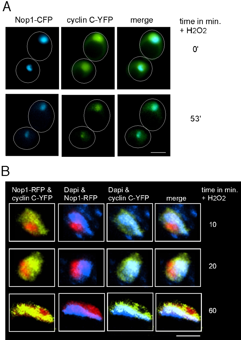
FIGURE 4: Cyclin C transits through the nucleolus in response to stress.
(A) Live cell imaging of immobilized wild type cells (FLY1589) expressing cyclin C-YFP and Nop1-CFP before (0 min) and following (53 min) exposure to 1.0 mM H2O2. Arrows indicate restricted cyclin C-YFP signal coinciding with Nop1-CFP.
(B) Enlarged fixed cell images of nucleus and surrounding area of wild type cells (RSY10) expressing cyclin C-YFP and Nop1-RFP. Cells were harvested and stained with DAPI following 1.2 mM H2O2 at the time points indicated.

