Table of contents
Volume 2, Issue 10, pp. 356 - 411, October 2015
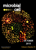
Cover: Fluorescent microphotography of a wild type yeast diploid strain and of several deletants of genes involved in the bridge-induced translocation regulatory pathway. The cells were stained with FUN-1 dye (6uM) and visualized with fluorescein and rhodamin filters. Inactive/dead cells appear as yellow while metabolically active/alive cells show red intravacuolar structures. Image by Valentina Tosato (International Center for Genetic Engineering and Biotechnology, Trieste, Italy); modified by MIC. The cover is published under the Creative Commons Attribution (CC BY) license.
Enlarge issue cover
Elongation factor-P at the crossroads of the host-endosymbiont interface
Andrei Rajkovic, Anne Witzky, William Navarre, Andrew J. Darwin and Michael Ibba
News and thoughts |
page 360-362 | 10.15698/mic2015.10.232 | Full text | PDF |
Abstract
Elongation factor P (EF-P) is an ancient bacterial translational factor that aids the ribosome in polymerizing oligo-prolines. EF-P structurally resembles tRNA and binds in-between the exit and peptidyl sites of the ribosome to accelerate the intrinsically slow reaction of peptidyl-prolyl bond formation. Recent studies have identified in separate organisms, two evolutionarily convergent EF-P post-translational modification systems (EPMS), split predominantly between gammaproteobacteria, and betaproteobacteria. In both cases EF-P receives a post-translational modification, critical for its function, on a highly conserved residue that protrudes into the peptidyl-transfer center of the ribosome. EPMSs are comprised of a gene(s) that synthesizes the precursor molecule used in modifying EF-P, and a gene(s) encoding an enzyme that reacts with the precursor molecule to catalyze covalent attachment to EF-P. However, not all organisms genetically encode a complete EPMS. For instance, some symbiotic bacteria harbor efp and the corresponding gene that enzymatically attaches the modification, but lack the ability to synthesize the substrate used in the modification reaction. Here we highlight the recent discoveries made regarding EPMSs, with a focus on how these incomplete modification pathways shape or have been shaped by the endosymbiont-host relationship.
Per aspera ad astra: When harmful chromosomal translocations become a plus value in genetic evolution. Lessons from Saccharomyces cerevisiae
Valentina Tosato and Carlo V. Bruschi
Reviews |
page 363-375 | 10.15698/mic2015.10.230 | Full text | PDF |
Abstract
In this review we will focus on chromosomal translocations (either spontaneous or induced) in budding yeast. Indeed, very few organisms tolerate so well aneuploidy like Saccharomyces, allowing in depth studies on chromosomal numerical aberrations. Many wild type strains naturally develop chromosomal rearrangements while adapting to different environmental conditions. Translocations, in particular, are valuable not only because they naturally drive species evolution, but because they might allow the artificial generation of new strains that can be optimized for industrial purposes. In this area, several methodologies to artificially trigger chromosomal translocations have been conceived in the past years, such as the chromosomal fragmentation vector (CFV) technique, the Cre-loxP procedure, the FLP/FRT recombination method and, recently, the bridge – induced translocation (BIT) system. An overview of the methodologies to generate chromosomal translocations in yeast will be presented and discussed considering advantages and drawbacks of each technology, focusing in particular on the recent BIT system. Translocants are important for clinical studies because translocated yeast cells resemble cancer cells from morphological and physiological points of view and because the translocation event ensues in a transcriptional de-regulation with a subsequent multi-factorial genetic adaptation to new, selective environmental conditions. The phenomenon of post-translocational adaptation (PTA) is discussed, providing some new unpublished data and proposing the hypothesis that translocations may drive evolution through adaptive genetic selection.
DNA damage checkpoint adaptation genes are required for division of cells harbouring eroded telomeres
Sofiane Y. Mersaoui, Serge Gravel, Victor Karpov, and Raymund J. Wellinger
Research Articles |
page 394-405 | 10.15698/mic2015.10.229 | Full text | PDF |
Abstract
In budding yeast, telomerase and the Cdc13p protein are two key players acting to ensure telomere stability. In the absence of telomerase, cells eventually enter a growth arrest which only few can overcome via a conserved process; such cells are called survivors. Survivors rely on homologous recombination-dependent mechanisms for telomeric repeat addition. Previously, we showed that such survivor cells also manage to bypass the loss of the essential Cdc13p protein to give rise to Cdc13-independent (or cap-independent) strains. Here we show that Cdc13-independent cells grow with persistently recognized DNA damage, which does not however result in a checkpoint activation; thus no defect in cell cycle progression is detectable. The absence of checkpoint signalling rather is due to the accumulation of mutations in checkpoint genes such as RAD24 or MEC1. Importantly, our results also show that cells that have lost the ability to adapt to persistent DNA damage, also are very much impaired in generating cap-independent cells. Altogether, these results show that while the capping process can be flexible, it takes a very specific genetic setup to allow a change from canonical capping to alternative capping. We hypothesize that in the alternative capping mode, genome integrity mechanisms are abrogated, which could cause increased mutation frequencies. These results from yeast have clear parallels in transformed human cancer cells and offer deeper insights into processes operating in pre-cancerous human cells that harbour eroded telomeres.
Formyl-methionine as a degradation signal at the N-termini of bacterial proteins
Konstantin I. Piatkov, Tri T. M. Vu, Cheol-Sang Hwang and Alexander Varshavsky
Research Articles |
page 376-393 | 10.15698/mic2015.10.231 | Full text | PDF |
Abstract
In bacteria, all nascent proteins bear the pretranslationally formed N-terminal formyl-methionine (fMet) residue. The fMet residue is cotranslationally deformylated by a ribosome-associated deformylase. The formylation of N-terminal Met in bacterial proteins is not strictly essential for either translation or cell viability. Moreover, protein synthesis by the cytosolic ribosomes of eukaryotes does not involve the formylation of N-terminal Met. What, then, is the main biological function of this metabolically costly, transient, and not strictly essential modification of N‑terminal Met, and why has Met formylation not been eliminated during bacterial evolution? One possibility is that the similarity of the formyl and acetyl groups, their identical locations in N‑terminally formylated (Nt‑formylated) and Nt-acetylated proteins, and the recently discovered proteolytic function of Nt-acetylation in eukaryotes might also signify a proteolytic role of Nt‑formylation in bacteria. We addressed this hypothesis about fMet‑based degradation signals, termed fMet/N-degrons, using specific E. coli mutants, pulse-chase degradation assays, and protein reporters whose deformylation was altered, through site-directed mutagenesis, to be either rapid or relatively slow. Our findings strongly suggest that the formylated N-terminal fMet can act as a degradation signal, largely a cotranslational one. One likely function of fMet/N-degrons is the control of protein quality. In bacteria, the rate of polypeptide chain elongation is nearly an order of magnitude higher than in eukaryotes. We suggest that the faster emergence of nascent proteins from bacterial ribosomes is one mechanistic and evolutionary reason for the pretranslational design of bacterial fMet/N‑degrons, in contrast to the cotranslational design of analogous Ac/N‑degrons in eukaryotes.
A bacterial volatile signal for biofilm formation
Yun Chen, Kevin Gozzi, and Yunrong Chai
Microreviews |
page 406-408 | 10.15698/mic2015.10.233 | Full text | PDF |
Abstract
Bacteria constantly monitor the environment they reside in and respond to potential changes in the environment through a variety of signal sensing and transduction mechanisms in a timely fashion. Those signaling mechanisms often involve application of small, diffusible chemical molecules. Volatiles are a group of small air-transmittable chemicals that are produced universally by all kingdoms of organisms. Past studies have shown that volatiles can function as cell-cell communication signals not only within species, but also cross-species. However, little is known about how the volatile-mediated signaling mechanism works. In our recent study (Chen, et al. mBio (2015), 6: e00392-15), we demonstrated that the soil bacterium Bacillus subtilis uses acetic acid as a volatile signal to coordinate the timing of biofilm formation within physically separated cells in the community. We also showed that the bacterium possesses an intertwined gene network to produce, secrete, sense, and respond to acetic acid, in stimulating biofilm formation. Interestingly, many of those genes are highly conserved in other bacterial species, raising the possibility that acetic acid may act as a volatile signal for cross-species communication.
The great escape: Pseudomonas breaks out of the lung
Angelica Zhang, Stephanie M. Rangel, and Alan R. Hauser
Microreviews |
page 409-411 | 10.15698/mic2015.10.234 | Full text | PDF |
Abstract
The Gram-negative bacterium Pseudomonas aeruginosa is a major cause of hospital-acquired infections and the focus of much attention due to its resistance to many conventional antibiotics. It harbors a wide range of disease-promoting virulence factors, including a type III secretion system. Here we review our recent study of ExoS, one of the effector proteins exported by this type III secretion system. Using a mouse model of pneumonia, we showed that the ADP-ribosyltransferase (ADPRT) activity of ExoS caused formation of “fields of cell injection” (FOCI) in the lungs. These FOCI represented ExoS-injected clusters of type I pneumocytes that became compromised, leading to disruption of the pulmonary-vascular barrier and subsequent bacterial dissemination from the lungs to the bloodstream. We discuss the potential mechanisms by which these processes occur as well as the novel techniques used to study ExoS function in vivo.


