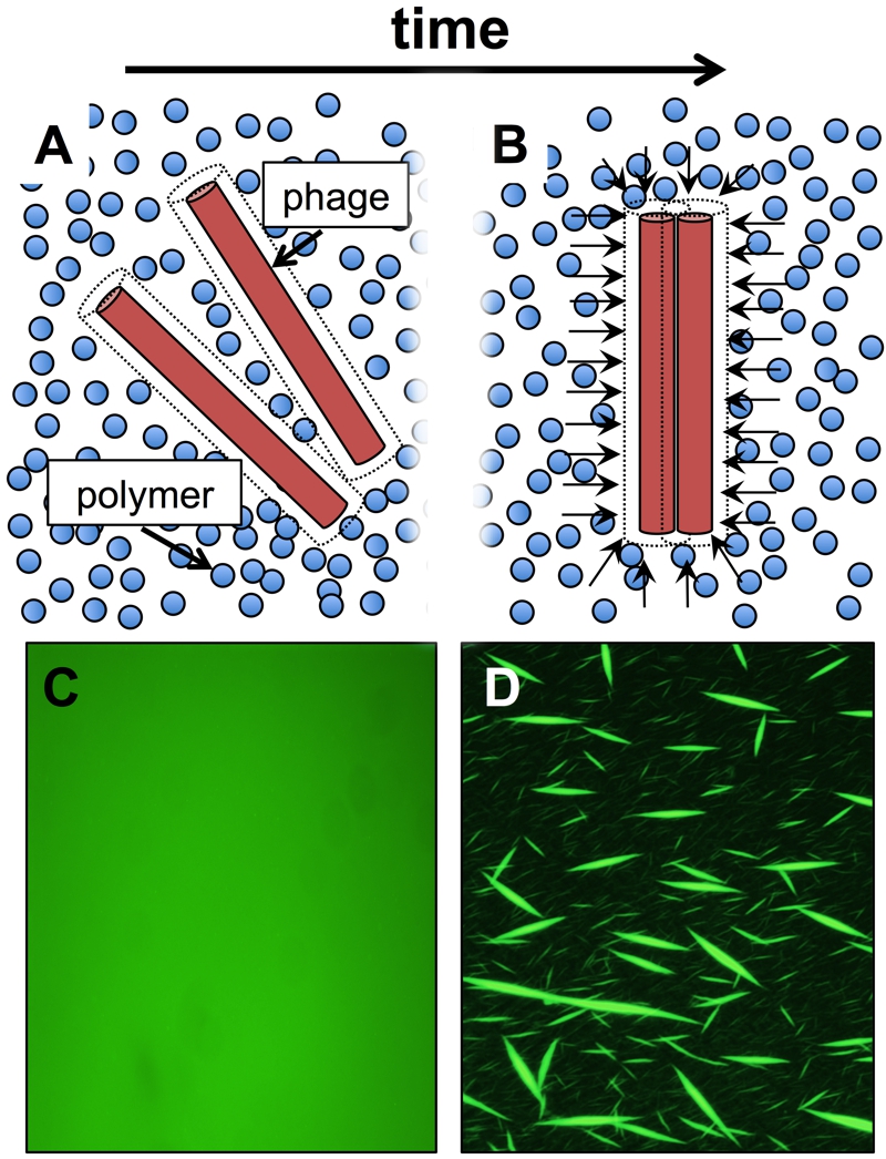
FIGURE 1: Schematic of how depletion attraction drives liquid crystal assembly from Pf phage within polymer-rich environments.
(A) Dashed lines around phage particles represent an exclusion volume not accessible to the polymer coil’s center of mass. When phages are dispersed, the volume inaccessible to the polymers is maximized.
(B) When phages come into close proximity to each other, their exclusion volumes overlap, increasing the total volume accessible to the polymers. This maximizes the entropy of the system. Arrows represent an osmotic imbalance that polymers exert on the liquid crystalline phage bundle. This osmotic pressure is what physically holds the liquid crystal together.
(C) Fluorescently labeled Pf phages (green) are initially dispersed when mixed with the host polymer hyaluronan.
(D) As time progresses, the phages form liquid crystalline structures.

