FIGURE 3:
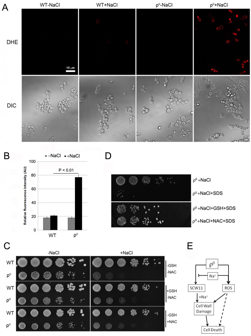
(A) and (B) Reactive oxygen species (ROS) production in the WT and ρ0 cells treated with 0.6 M NaCl for 15 min.
(A) After salt stress, ρ0 cells displayed a higher fluorescence level than WT cells. Staining with 5 µM dihydroethidium (DHE) was used.
(B) Quantification of ROS production. Relative fluorescence intensities were measured by the ImageJ software. Values presented are means of three independent experiments. About 300 cells were measured during each experiment.
(C) Antioxidant GSH or NAC slightly decreases the salt sensitivity of ρ0 cells. Log phase cells were spotted directly onto YPD plates containing 0.6 M NaCl and GSH (10 mM) or NAC (20 mM). Cells were cultured at 30οC for 2 days.
(D) Addition of antioxidant GSH or NAC reduced the sensitivity of ρ0 cells towards SDS after salt stress. Cells were treated with 0.6 M NaCl in presence of GSH (10 mM) or NAC (20 mM) for 1 hr. Cells were washed, treated with SDS for 0.5 hr and spotted on YPD plates. Cells were then cultured at 30οC for 2 days.
(E) Diagram showing the possible involvement of SCW11 and ROS in salt stress-induced cell wall damage and cell death of ρ0 cells.

