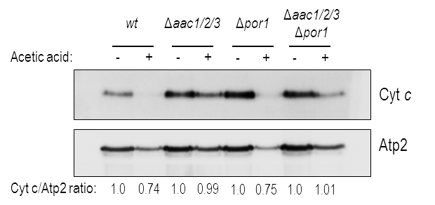
FIGURE 3: Cyt c is released from wt and Δpor1 mitochondria.
Cells were pre-cultured in YPD, and then grown O.N. in YPGal until an OD640nm of 1.5-2.0 was reached, before adding acetic acid. One representative experiment of cyt c immunodetection in wt, Δaac1/2/3, Δpor1, Δaac1/2/3Δpor1 mitochondrial fractions, isolated from control and acetic-treated cells, is presented. The beta subunit of the F1 sector of mitochondrial FOF1 ATP synthase (Atp2p) was used as control for the mitochondrial fractions. A densitometric analysis was performed (ImageJ software) and the corresponding cyt c / Atp2 protein ratios are presented.

