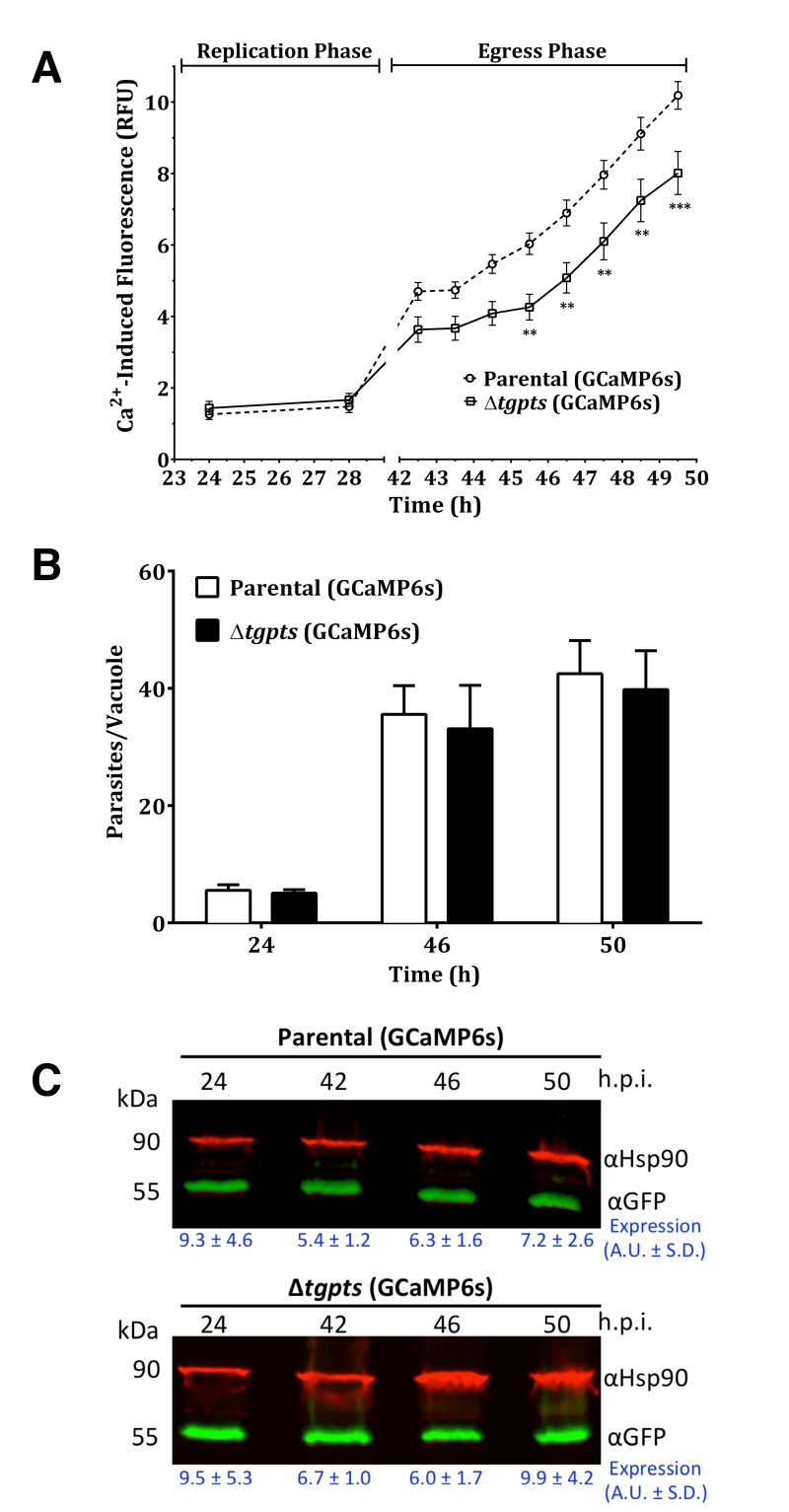
FIGURE 4: The PTS-knockout mutant exhibits a dysregulation of cytosolic calcium during its natural egression but not during the proliferation phase. (A) Quantitation of EGFP-derived fluorescence during replication and egress phases of GECI strains. Human fibroblast cultures were infected with GCaMP6s-transgenic parental or Δtgpts strains. Calcium-induced EGFP intensity was measured in living cultures using a microplate reader. Graphs show the mean ± SEM of 9 assays, each in tripli-cates (student’s t-test, **p< 0.01, ***p<0.001). (B) Replication rates of the parental and mutant strains expressing GECI. Intracellular parasites replicating in their vacuoles were quantified at specified time points (representing replication, early and late egress) after immunostaining with anti-TgGap45 antibody (mean ± SEM, n=3 assays). Consistent with parasite counts, EGFP signal during the replication phase was indistinguishable in the two GCaMP6s-transgenic strains. (C) A representative western blot depicting the expression of M13-CpEGFP-CaM in the two parasite strains. TgHsp90 was used for normalizing the expression of GCaMP6s in each lane.
By continuing to use the site, you agree to the use of cookies. more information
The cookie settings on this website are set to "allow cookies" to give you the best browsing experience possible. If you continue to use this website without changing your cookie settings or you click "Accept" below then you are consenting to this. Please refer to our "privacy statement" and our "terms of use" for further information.

