Table of contents
Volume 7, Issue 4, pp. 93 - 118, April 2020
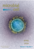
Cover: Transmission electron micrograph of a SARS-CoV-2 virus particle, isolated from a patient (image captured and color-enhanced at the National Institute of Allergy and Infectious Diseases, National Institutes of Health (USA) Integrated Research Facility (IRF); the image was modified by MIC). The cover is published under the Creative Commons Attribution (CC BY) license.
Enlarge issue cover
Regulation of anti-microbial autophagy by factors of the complement system
Christophe Viret, Aurore Rozières, Rémi Duclaux-Loras, Gilles Boschetti, Stéphane Nancey and Mathias Faure
Reviews |
page 93-105 | 10.15698/mic2020.04.712 | Full text | PDF |
Abstract
The complement system is a major component of innate immunity that participates in the defense of the host against a myriad of pathogenic microorganisms. Activation of complement allows for both local inflammatory response and physical elimination of microbes through phagocytosis or lysis. The system is highly efficient and is therefore finely regulated. In addition to these well-established properties, recent works have revealed that components of the complement system can be involved in a variety of other functions including in autophagy, the conserved mechanism that allows for the targeting and degradation of cytosolic materials by the lysosomal pathway after confining them into specialized organelles called autophagosomes. Besides impacting cell death, development or metabolism, the complement factors-autophagy connection can greatly modulate the cell autonomous, anti-microbial activity of autophagy: xenophagy. Both surface receptor-ligand interactions and intracellular interactions are involved in the modulation of the autophagic response to intracellular microbes by complement factors. Here, we review works that relate to the recently discovered connections between factors of the complement system and the functioning of autophagy in the context of host-pathogen relationship.
Stable and destabilized GFP reporters to monitor calcineurin activity in Saccharomyces cerevisiae
Jutta Diessl, Arpita Nandy, Christina Schug, Lukas Habernig and Sabrina Büttner
Research Articles |
page 106-114 | 10.15698/mic2020.04.713 | Full text | PDF |
Abstract
The protein phosphatase calcineurin is activated in response to rising intracellular Ca2+ levels and impacts fundamental cellular processes in organisms ranging from yeast to humans. In fungi, calcineurin orchestrates cellular adaptation to diverse environmental challenges and is essential for virulence of pathogenic species. To enable rapid and large-scale assessment of calcineurin activity in living, unperturbed yeast cells, we have generated stable and destabilized GFP transcriptional reporters under the control of a calcineurin-dependent response element (CDRE). Using the reporters, we show that the rapid dynamics of calcineurin activation and deactivation can be followed by flow cytometry and fluorescence microscopy. This system is compatible with live/dead staining that excludes confounding dead cells from the analysis. The reporters provide technology to monitor calcineurin dynamics during stress and ageing and may serve as a drug-screening platform to identify novel antifungal compounds that selectively target calcineurin.
More than flipping the lid: Cdc50 contributes to echinocandin resistance by regulating calcium homeostasis in Cryptococcus neoformans
Chengjun Cao and Chaoyang Xue
Microreviews |
page 115-118 | 10.15698/mic2020.04.714 | Full text | PDF |
Abstract
Echinocandins are the newest fungicidal drug class approved for clinical use against common invasive mycoses. Yet, they are ineffective against cryptococcosis, predominantly caused by Cryptococcus neoformans. The underlying mechanisms of innate echinocandin resistance in C. neoformans remain unclear. We know that Cdc50, the β-subunit of the lipid translocase (flippase), mediates echinocandin resistance, as loss of the CDC50 gene sensitizes C. neoformans to caspofungin, a member of the echinocandins class. We sought to elucidate how Cdc50 facilitates caspofungin resistance by performing a forward genetic screen for cdc50Δ suppressor mutations that are caspofungin resistant. We identified a novel mechanosensitive calcium channel protein Crm1 that correlates with Cdc50 function (Cao et al., 2019). In addition to regulating phospholipid translocation, Cdc50 also interacts with Crm1 to regulate intracellular calcium homeostasis and calcium/calcineurin signaling that likely drives caspofungin resistance in C. neoformans. Our study revealed a novel dual function of Cdc50 that connects lipid flippase with calcium signaling. These unexpected findings provide new insights into the mechanisms of echinocandin resistance in C. neoformans that may drive future drug design.


