Back to issue: December 2015
Microbial Cell – December 2015
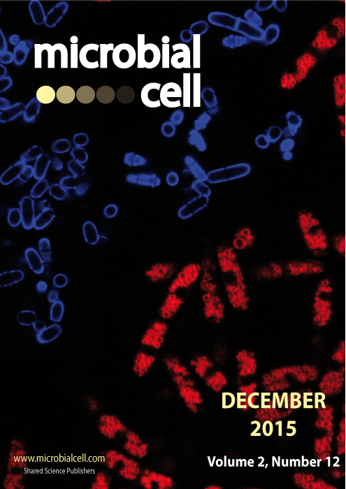
Schizosaccharomyces pombe cells stained with calcofluor white (blue) to visualize the morphology of the whole cell or with the vital dye FM4-64 (red) to visualize the vacuoles. Image by Michael Bond and Sara Mole (University College London, UK); modified by MIC. The cover is published under the Creative Commons Attribution (CC BY) license.

