Table of contents
Volume 1, Issue 2, pp. 51 - 69, February 2014
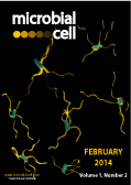
Cover: Motile Salmonella with long helical flagella assembled on their cell surface. Flagella grow to several times the length of the bacterium. Images acquired using epifluorescence microscopy. Pictures by Lewis D. B. Evans, Colin Hughes and Gillian M. Fraser (University of Cambridge). The cover is published under the Creative Commons Attribution-NonCommercial-ShareAlike 3.0 license.
Enlarge issue cover
Mitochondrial protein import under kinase surveillance
Magdalena Opalińska and Chris Meisinger
In a nutshell |
page 51-57 | 10.15698/mic2014.01.127 | Full text | PDF |
Abstract
Despite the simplicity of the yeast Saccharomyces cerevisiae, its basic cellular machinery tremendously mirrors that of higher eukaryotic counterparts. Thus, this unicellular organism turned out to be an invaluable model system to study the countless mechanisms that govern life of the cell. Recently, it has also enabled the deciphering of signalling pathways that control flux of mitochondrial proteins to the organelle according to metabolic requirements. For decades mitochondria were considered autonomous organelles that are only partially incorporated into cellular signalling networks. Consequently, only little has been known about the role of reversible phosphorylation as a meaningful mechanism that orchestrates mitochondrial biology accordingly to cellular needs. Therefore, research in this direction has been vastly neglected. However, findings over the past few years have changed this view and new exciting fields in mitochondrial biology have emerged. Here, we summarize recent discoveries in the yeast model system that point towards a vital role of reversible phosphorylation in regulation of mitochondrial protein import.
Deletion of AIF1 but not of YCA1/MCA1 protects Saccharomyces cerevisiae and Candida albicans cells from caspofungin-induced programmed cell death
Christopher Chin, Faith Donaghey, Katherine Helming, Morgan McCarthy, Stephen Rogers, and Nicanor Austriaco.
Research Reports |
page 58-63 | 10.15698/mic2014.01.119 | Full text | PDF |
Abstract
Caspofungin was the first member of a new class of antifungals called echinocandins to be approved by a drug regulatory authority. Like the other echinocandins, caspofungin blocks the synthesis of β(1,3)-D-glucan of the fungal cell wall by inhibiting the enzyme, β(1,3)-D-glucan synthase. Loss of β(1,3)-D-glucan leads to osmotic instability and cell death. However, the precise mechanism of cell death associated with the cytotoxicity of caspofungin was unclear. We now provide evidence that Saccharomyces cerevisiae cells cultured in media containing caspofungin manifest the classical hallmarks of programmed cell death (PCD) in yeast, including the generation of reactive oxygen species (ROS), the fragmentation of mitochondria, and the production of DNA strand breaks. Our data also suggests that deleting AIF1 but not YCA1/MCA1 protects S. cerevisiae and Candida albicans from caspofungin-induced cell death. This is not only the first time that AIF1 has been specifically tied to cell death in Candida but also the first time that caspofungin resistance has been linked to the cell death machinery in yeast.
Building a flagellum in biological outer space
Lewis D. B. Evans, Colin Hughes and Gillian M. Fraser
Microreviews |
page 64-66 | 10.15698/mic2014.01.128 | Full text | PDF |
Abstract
Flagella, the rotary propellers on the surface of bacteria, present a paradigm for how cells build and operate complex molecular ‘nanomachines’. Flagella grow at a constant rate to extend several times the length of the cell, and this is achieved by thousands of secreted structural subunits transiting through a central channel in the lengthening flagellum to incorporate into the nascent structure at the distant extending tip. A great mystery has been how flagella can assemble far outside the cell where there is no conventional energy supply to fuel their growth. Recent work published by Evans et al. [Nature (2013) 504: 287-290], has gone some way towards solving this puzzle, presenting a simple and elegant transit mechanism in which growth is powered by the subunits themselves as they link head-to-tail in a chain that is pulled through the length of the growing structure to the tip. This new mechanism answers an old question and may have resonance in other assembly processes.
Intersubunit communications within KaiC hexamers contribute the robust rhythmicity of the cyanobacterial circadian clock
Yohko Kitayama, Taeko Nishiwaki-Ohkawa, Takao Kondo
Microreviews |
page 67-69 | 10.15698/mic2014.01.129 | Full text | PDF |
Abstract
Circadian rhythms, endogenous oscillations with periods of ~24 h, are found in many organisms, and they enhance fitness in alternating day/night environments. In cyanobacteria, three clock proteins, KaiA, KaiB, and KaiC, control the timekeeping mechanism. KaiC, the central component of the system, is a hexameric ATPase that also has autokinase and autophosphatase activities. It has been assumed that KaiC’s hexameric structure was critical for regulation of the circadian clock; however, the underlying molecular mechanism of such regulation has remained unclear. Recently, we elucidated the regulation of KaiC’s activities by its phosphorylation state, in the context of its hexameric structure. We found that local interactions at subunit interfaces regulate KaiC’s activities by coupling the nucleotide-binding states. We also discovered the mechanism of regulation by intersubunit communication in KaiC hexamers. Our observations suggest that intersubunit communication precisely synchronizes KaiC subunits to avoid dephasing, and contributes to the robustness of circadian rhythms in cyanobacteria [Kitayama, Y. et al. Nat. Commun. 4:2897 doi: 10.1038/ncomms3897 (2013)].


