Back to issue: February 2021
Microbial Cell February 2021
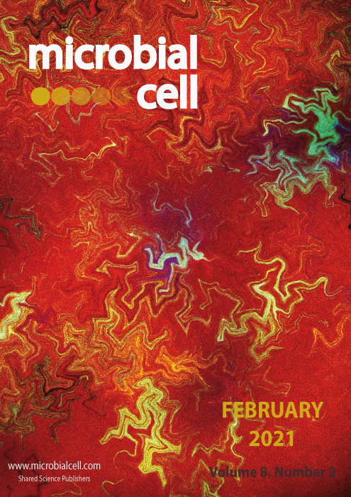
The image was obtained by confocal microscopy (using a Leica SP5). It consists of Bacillus subtilis transformed (separately) with fluorescent proteins TagRFP-T, sfGFP, TagBFP, mKate2 and mOrange2, mixed and plated on solid media for 24h. Images were obtained directly from petri dishes by positioning a cover slip over the growing cells (image by Fernan Federici, Pontificia Universidad Catolica de Chile (Chile) and iBio Institute; Tim Rudge, PJ Steiner and Jim Haseloff, University of Cambridge (UK); image retrieved via wellcomecollection.org); image modified by MIC. The cover is published under the Creative Commons Attribution (CC BY) license.

