Table of contents
Volume 7, Issue 1, pp. 1 - 31, January 2020
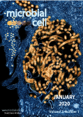
Cover: Colorized transmission electron micrograph of Ebola virus nucleocapsids (small orange circles) and virus particles (larger orange filamentous forms) within infected African green monkey kidney cells (image by the National Institute of Allergy and Infectious Diseases (USA); retrieved via Flickr; the image was modified by MIC). The cover is published under the Creative Commons Attribution (CC BY) license.
Enlarge issue cover
The role of Lactobacillus species in the control of Candida via biotrophic interactions
Isabella Zangl, Ildiko-Julia Pap, Christoph Aspöck and Christoph Schüller
Reviews |
page 1-14 | 10.15698/mic2020.01.702 | Full text | PDF |
Abstract
Microbial communities have an important role in health and disease. Candida spp. are ubiquitous commensals and sometimes opportunistic fungal pathogens of humans, colonizing mucosal surfaces of the genital, urinary, respiratory and gastrointestinal tracts and the oral cavity. They mainly cause local mucosal infections in immune competent individuals. However, in the case of an ineffective immune defense, Candida infections may become a serious threat. Lactobacillus spp. are part of the human microbiome and are natural competitors of Candida in the vaginal environment. Lactic acid, low pH and other secreted metabolites are environmental signals sensed by fungal species present in the microbiome. This review briefly discusses the ternary interaction between host, Lactobacillus species and Candida with regard to fungal infections and the potential antifungal and fungistatic effect of Lactobacillus species. Our understanding of these interactions is incomplete due to the variability of the involved species and isolates and the complexity of the human host.
Yeast can express and assemble bacterial secretins in the mitochondrial outer membrane
Janani Natarajan, Anasuya Moitra, Sussanne Zabel, Nidhi Singh, Samuel Wagner and Doron Rapaport
Research Articles |
page 15-27 | 10.15698/mic2020.01.703 | Full text | PDF |
Abstract
Secretins form large multimeric pores in the outer membrane (OM) of Gram-negative bacteria. These pores are part of type II and III secretion systems (T2SS and T3SS, respectively) and are crucial for pathogenicity. Recent structural studies indicate that secretins form a structure rich in β-strands. However, little is known about the mechanism by which secretins assemble into the OM. Based on the conservation of the biogenesis of β-barrel proteins in bacteria and mitochondria, we used yeast cells as a model system to study the assembly process of secretins. To that end, we analyzed the biogenesis of PulD (T2SS), SsaC (T3SS) and InvG (T3SS) in wild type cells or in cells mutated for known mitochondrial import and assembly factors. Our results suggest that secretins can be expressed in yeast cells, where they are enriched in the mitochondrial fraction. Interestingly, deletion of mitochondrial import receptors like Tom20 and Tom70 reduces the mitochondrial association of PulD but does not affect that of InvG. SsaC shows another dependency pattern and its membrane assembly is enhanced by the absence of Tom70 and compromised in cells lacking Tom20 or the topogenesis of outer membrane β-barrel proteins (TOB) complex component, Mas37. Collectively, these findings suggest that various secretins can follow different pathways to assemble into the bacterial OM.
A holobiont view on thrombosis: unravelling the microbiota’s influence on arterial thrombus growth
Giulia Pontarollo, Klytaimnistra Kiouptsi and Christoph Reinhardt
Microreviews |
page 28-31 | 10.15698/mic2020.01.704 | Full text | PDF |
Abstract
The commensal microbiota has co-evolved with its host, colonizing all body surfaces. Therefore, this microbial ecosystem is intertwined with host physiology at multiple levels. While it is evident that microbes that reach the blood stream can trigger thrombus formation, it remains poorly explored if the wealth of microbes that colonize the body surfaces of the mammalian host can be regarded as a modifier of cardiovascular disease (CVD) development. To experimentally address the microbiota’s role in the development of atherosclerotic lesions and arterial thrombosis, we generated a germ-free (GF) low-density lipoprotein receptor-deficient (Ldlr-/-) atherosclerosis mouse model (Kiouptsi et al., mBio, 2019) and explored the role of nutritional composition on arterial thrombogenesis.


