Table of contents
Volume 4, Issue 3, pp. 69 - 107, March 2017
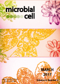
Cover: Combination of (i) fluorescense images of intracellular
Toxoplasma gondii tachyzoites showing division process by endodyogeny. Parasites were stained with anti-GAP45 for mother cell pellicle, anti-IMC1 for daughter and mother cell pellicle and DAPI for nucleus and (ii) an electron micrograph of an intracellular rosette of
Toxoplasma gondii (image by Erica dos Santos Martins-Duarte and Wanderley de Souza, Universidade Federal do Rio de Janeiro, Brazil); image modified by MIC. The cover is published under the Creative Commons Attribution (CC BY) license.
Enlarge issue cover
Transceptors as a functional link of transporters and receptors
George Diallinas
Editorial |
page 69-73 | 10.15698/mic2017.03.560 | Full text | PDF |
Abstract
Cells need to communicate with their environment in order to obtain nutrients, grow, divide and respond to signals related to adaptation in changing physiological conditions or stress. A very basic question in biology is how cells, especially of those organisms living in rapidly changing habitats, sense their environment. Apparently, this question is of particular importance to all free-living microorganisms. The critical role of receptors, transporters and channels, transmembrane proteins located in the plasma membrane of all types of cells, in signaling environmental changes is well established. A relative newcomer in environment sensing are the so called transceptors, membrane proteins that possess both solute transport and receptor-like signaling activities. Now, the transceptor concept is further enlarged to include micronutrient sensing via the iron and zinc high-affinity transporters of Saccharomyces cerevisiae. Interestingly, what seems to underline the transport and/or sensing function of receptors, transporters and transceptors is ligand-induced conformational alterations recognized by downstream intracellular effectors.
Identification of Ftr1 and Zrt1 as iron and zinc micronutrient transceptors for activation of the PKA pathway in Saccharomyces cerevisiae
Joep Schothorst, Griet Van Zeebroeck, Johan M. Thevelein
Research Articles |
page 74-89 | 10.15698/mic2017.03.561 | Full text | PDF |
Abstract
Multiple types of nutrient transceptors, membrane proteins that combine a transporter and receptor function, have now been established in a variety of organisms. However, so far all established transceptors utilize one of the macronutrients, glucose, amino acids, ammonium, nitrate, phosphate or sulfate, as substrate. This is also true for the Saccharomyces cerevisiae transceptors mediating activation of the PKA pathway upon re-addition of a macronutrient to glucose-repressed cells starved for that nutrient, re-establishing a fermentable growth medium. We now show that the yeast high-affinity iron transporter Ftr1 and high-affinity zinc transporter Zrt1 function as transceptors for the micronutrients iron and zinc. We show that replenishment of iron to iron-starved cells or zinc to zinc-starved cells triggers within 1-2 minutes a rapid surge in trehalase activity, a well-established PKA target. The activation with iron is dependent on Ftr1 and with zinc on Zrt1, and we show that it is independent of intracellular iron and zinc levels. Similar to the transceptors for macronutrients, Ftr1 and Zrt1 are strongly induced upon iron and zinc starvation, respectively, and they are rapidly downregulated by substrate-induced endocytosis. Our results suggest that transceptor-mediated signaling to the PKA pathway may occur in all cases where glucose-repressed yeast cells have been starved first for an essential nutrient, causing arrest of growth and low activity of the PKA pathway, and subsequently replenished with the lacking nutrient to re-establish a fermentable growth medium. The broadness of the phenomenon also makes it likely that nutrient transceptors use a common mechanism for signaling to the PKA pathway.
A multigene family encoding surface glycoproteins in Trypanosoma congolense
Magali Thonnus, Amandine Guérin, Loïc Rivière
Research Reports |
page 90-97 | 10.15698/mic2017.03.562 | Full text | PDF |
Abstract
Trypanosoma congolense, the causative agent of the most important livestock disease in Africa, expresses specific surface proteins involved in its parasitic lifestyle. Unfortunately, the complete repertoire of such molecules is far from being deciphered. As these membrane components are exposed to the host environment, they could be used as therapeutic or diagnostic targets. By mining the T. congolense genome database, we identified a novel family of lectin-like glycoproteins (TcoClecs). These molecules are predicted to have a transmembrane domain, a tandem repeat amino acid motif, a signal peptide and a C-type lectin-like domain (CTLD). This paper depicts several experimental arguments in favor of a surface localization in bloodstream forms of T. congolense. A TcoClec gene was heterologously expressed in U-2 OS cells and the product could be partially found at the plasma membrane. TcoClecs were also localized at the surface of T. congolense bloodstream forms. The signal was suppressed when the cells were treated with a detergent to remove the plasma membrane or with trypsin to « shave » the parasites and remove their external proteins. This suggests that TcoClecs could be potential diagnostic or therapeutic antigens of African animal trypanosomiasis. The potential role of these proteins in T. congolense as well as in other trypanosomatids is discussed.
Chlamydia trachomatis’ struggle to keep its host alive
Barbara S. Sixt, Raphael H. Valdivia, Guido Kroemer
Microreviews |
page 101-104 | 10.15698/mic2017.03.564 | Full text | PDF |
Abstract
Bacteria of the phylum Chlamydiae infect a diverse range of eukaryotic host species, including vertebrate animals, invertebrates, and even protozoa. Characteristics shared by all Chlamydiae include their obligate intracellular lifestyle and a biphasic developmental cycle. The infectious form, the elementary body (EB), invades a host cell and differentiates into the replicative form, the reticulate body (RB), which proliferates within a membrane-bound compartment, the inclusion. After several rounds of division, RBs retro-differentiate into EBs that are then released to infect neighboring cells. The consequence of this obligatory transition between replicative and infectious forms inside cells is that Chlamydiae absolutely depend on the viability and functionality of their host cell throughout the entire infection cycle. We recently conducted a forward genetic screen in Chlamydia trachomatis, a common sexually transmitted human pathogen, and identified a mutant that caused premature death in the majority of infected host cells. We employed emerging genetic tools in Chlamydia to link this cytotoxicity to the loss of the protein CpoS (Chlamydia promoter of survival) that normally localizes to the membrane of the pathogen-containing vacuole. CpoS-deficient bacteria also induced an exaggerated type-1 interferon response in infected cells, produced reduced numbers of infectious EBs in cell culture, and were cleared faster from the mouse genital tract in a transcervical infection model in vivo. The analysis of this CpoS-deficient mutant yielded unique insights into the nature of cell-autonomous defense responses against Chlamydia and highlighted the importance of Chlamydia-mediated control of host cell fate for the success of the pathogen.
Advancing host-directed therapy for tuberculosis: new therapeutic insights from the Toxoplasma gondii
Chul-Su Yang
Microreviews |
page 105-107 | 10.15698/mic2017.03.565 | Full text | PDF |
Abstract
Tuberculosis (TB) drug-development strategies, a wide range of candidate host-directed therapies (HDT)s-including new and repurposed drugs, biologics, and cellular therapies-have been proposed to accelerate eradication of infection and overcome the problems associated with current treatment regimens. By investigating the interaction between macrophages and the intracellular parasite Toxoplasma gondii (T. gondii), we uncovered that infection-induced signaling pathways suggest possibilities for the development of novel therapeutic modalities for TB that target the intracellular signaling pathways permitting the replication of Mycobacterium tuberculosis (MTB).
New insights into the function of a versatile class of membrane molecular motors from studies of Myxococcus xanthus surface (gliding) motility
Tâm Mignot, Marcelo Nöllmann
Microreviews |
page 98-100 | 10.15698/mic2017.03.563 | Full text | PDF |
Abstract
Cell motility is a central function of living cells, as it empowers colonization of new environmental niches, cooperation, and development of multicellular organisms. This process is achieved by complex yet precise energy-consuming machineries in both eukaryotes and bacteria. Bacteria move on surfaces using extracellular appendages such as flagella and pili but also by a less-understood process called gliding motility. During this process, rod-shaped bacteria move smoothly along their long axis without any visible morphological changes besides occasional bending. For this reason, the molecular mechanism of gliding motility and its origin have long remained a complete mystery. An important breakthrough in the understanding of gliding motility came from single cell and genetic studies in the delta-proteobacterium Myxococcus xanthus. These early studies revealed, for the first time, the existence of bacterial Focal Adhesion complexes (FA). FAs are formed at the bacterial pole and rapidly move towards the opposite cell pole. Their attachment to the underlying surface is linked to cell propulsion, in a process similar to the rearward translocation of actomyosin complexes in Apicomplexans. The protein machinery that forms at FAs was shown to contain up to seventeen proteins predicted to localize in all layers of the bacterial cell envelope, the cytosolic face, the inner membrane (IM), the periplasmic space and the outer membrane (OM). Among these proteins, a proton-gated channel at the inner membrane was identified as the molecular motor. Thus, thrust generation requires the transduction of traction forces generated at the inner membrane through the cell envelope beyond the rigid barrier of the bacterial peptidoglycan.


