
Back to issue: May 2014
Microbial Cell – May 2014
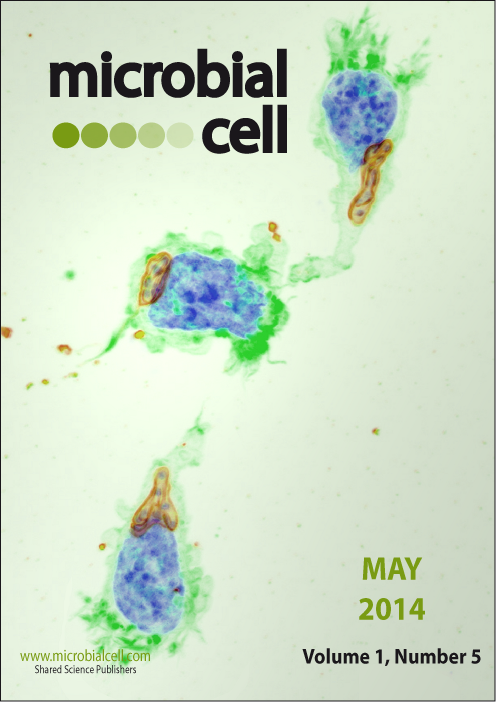
Bovine macrophages infected with the intracellular, protozoan parasite Theileria annulata migrating inside matrigel. Leading edge is rich in F-actin (green) and dynamically remodeled. Parasite in red is located behind host cell nucleus (blue) with respect to the direction of migration. Surpass image of 15 individual confocal sections is shown. Green: F-actin (phalloidin Alexa488), Red: parasite (anti-Theileria annulata surface protein TASP, Cy3), Blue: DNA (hoechst). Picture by Martin Baumgartner (University of Zürich, Switzerland). The cover is published under the Creative Commons Attribution (CC BY) license.