Table of contents
Volume 4, Issue 5, pp. 140 - 181, May 2017
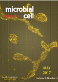
Cover: Colorized photographic detail of a haemocytometer with propidium iodide-stained
Saccharomyces cerevisiae cells (image by Andreas Zimmermann, Didac Carmona-Gutierrez and Frank Madeo, University of Graz, Austria). The cover is published under the Creative Commons Attribution (CC BY) license.
Enlarge issue cover
When a ribosomal protein grows up – the ribosome assembly path of Rps3
Brigitte Pertschy
News and thoughts |
page 140-143 | 10.15698/mic2017.05.571 | Full text | PDF |
Abstract
The biogenesis of ribosomes is a central process in all dividing cells. Eukaryotic ribosomes are composed of a large 60S and a small 40S subunit, each comprising a complex assembly of ribosomal RNA (rRNA) and ribosomal proteins (r-proteins). The synthesis of these constituents is spatially separated, with r-proteins being produced by translation in the cytoplasm, while rRNA is generated by transcription in the nucleus. Hence, the arrangement of r-proteins and rRNA into large ribonucleoprotein complexes requires dedicated mechanisms ensuring their encounter in the same compartment. To this end, r-proteins need to be safely delivered to the nucleus where they assemble with the rRNA. Beyond these initial challenges, the synthesis of ribosomes does not merely comprise the joining of r-proteins with rRNA, but occurs in a complex assembly line involving multiple maturation steps, including the processing and folding of rRNA. R-proteins usually have composite rRNA binding sites, with several different rRNA helices contributing to the full interaction. Not all of these interaction sites may already be accessible at the point when an r-protein is incorporated, necessitating that some of the r-protein-rRNA contacts are formed at later maturation stages. In our two recent studies, we investigated the ribosome assembly path of r-proteins in the yeast Saccharomyces cerevisiae using the small subunit r-protein S3 (Rps3) as a model. Our studies revealed intricate mechanisms to protect the protein, transport it into the nucleus, integrate it into pre-ribosomal precursor particles and promote its final stable association with 40S subunits.
Placeholder factors in ribosome biogenesis: please, pave my way
Francisco J. Espinar-Marchena, Reyes Babiano and Jesús de la Cruz
Reviews |
page 144-168 | 10.15698/mic2017.05.572 | Full text | PDF |
Abstract
The synthesis of cytoplasmic eukaryotic ribosomes is an extraordinarily energy-demanding cellular activity that occurs progressively from the nucleolus to the cytoplasm. In the nucleolus, precursor rRNAs associate with a myriad of trans-acting factors and some ribosomal proteins to form pre-ribosomal particles. These factors include snoRNPs, nucleases, ATPases, GTPases, RNA helicases, and a vast list of proteins with no predicted enzymatic activity. Their coordinate activity orchestrates in a spatiotemporal manner the modification and processing of precursor rRNAs, the rearrangement reactions required for the formation of productive RNA folding intermediates, the ordered assembly of the ribosomal proteins, and the export of pre-ribosomal particles to the cytoplasm; thus, providing speed, directionality and accuracy to the overall process of formation of translation-competent ribosomes. Here, we review a particular class of trans-acting factors known as “placeholders”. Placeholder factors temporarily bind selected ribosomal sites until these have achieved a structural context that is appropriate for exchanging the placeholder with another site-specific binding factor. By this strategy, placeholders sterically prevent premature recruitment of subsequently binding factors, premature formation of structures, avoid possible folding traps, and act as molecular clocks that supervise the correct progression of pre-ribosomal particles into functional ribosomal subunits. We summarize the current understanding of those factors that delay the assembly of distinct ribosomal proteins or subsequently bind key sites in pre-ribosomal particles. We also discuss recurrent examples of RNA-protein and protein-protein mimicry between rRNAs and/or factors, which have clear functional implications for the ribosome biogenesis pathway.
A simple microfluidic platform to study age-dependent protein abundance and localization changes in Saccharomyces cerevisiae
Margarita Cabrera, Daniele Novarina, Irina L. Rempel, Liesbeth M. Veenhoff, and Michael Chang
Research Reports |
page 169-174 | 10.15698/mic2017.05.573 | Full text | PDF |
Abstract
The budding yeast Saccharomyces cerevisiae divides asymmetrically, with a smaller daughter cell emerging from its larger mother cell. While the daughter lineage is immortal, mother cells age with each cell division and have a finite lifespan. The replicative ageing of the yeast mother cell has been used as a model to study the ageing of mitotically active human cells. Several microfluidic platforms, which use fluid flow to selectively remove daughter cells, have recently been developed that can monitor cell physiology as mother cells age. However, these platforms are not trivial to set up and users often require many hours of training. In this study, we have developed a simple system, which combines a commercially available microfluidic platform (the CellASIC ONIX Microfluidic Platform) and a genetic tool to prevent the proliferation of daughter cells (the Mother Enrichment Program), to monitor protein abundance and localization changes during approximately the first half of the yeast replicative lifespan. We validated our system by observing known age-dependent changes, such as decreased Sir2 abundance, and have identified a protein with a previously unknown age-dependent change in localization.
Insights from the redefinition of Helicobacter pylori lipopolysaccharide O-antigen and core-oligosaccharide domains
Hong Li, Tiandi Yang, Tingting Liao, Aleksandra W. Debowski, Hans-Olof Nilsson, Stuart M. Haslam, Anne Dell, Keith A. Stubbs, Barry J. Marshall and Mohammed Benghezal
Microreviews |
page 175-178 | 10.15698/mic2017.05.574 | Full text | PDF |
Abstract
H. pylori is a Gram-negative extracellular bacterium, first discovered by the Australian physicians Barry Marshall and Robin Warren in 1982, that colonises the human stomach mucosa. It is the leading cause of peptic ulcer and commonly infects humans worldwide with prevalence as high as 90% in some countries. H. pylori infection usually results in asymptomatic chronic gastritis, however 10-15% of cases develop duodenal or gastric ulcers and 1-3% develop stomach cancer. Infection is generally acquired during childhood and persists for life in the absence of antibiotic treatment. H. pylori has had a long period of co-evolution with humans, going back to human migration out of Africa. This prolonged relationship is likely to have shaped the overall host-pathogen interactions and repertoire of virulence strategies which H. pylori employs to establish robust colonisation, escape immune responses and persist in the gastric niche. In this regard, H. pylori lipopolysaccharide (LPS) is a key surface determinant in establishing colonisation and persistence via host mimicry and resistance to cationic antimicrobial peptides. Thus, elucidation of the H. pylori LPS structure and corresponding biosynthetic pathway represents an important step towards better understanding of H. pylori pathogenesis and the development of novel therapeutic interventions.
Post-transcriptional regulation of ribosome biogenesis in yeast
Isabelle C. Kos-Braun and Martin Koš
Microreviews |
page 179-181 | 10.15698/mic2017.05.575 | Full text | PDF |
Abstract
Most microorganisms are exposed to the constantly and often rapidly changing environment. As such they evolved mechanisms to balance their metabolism and energy expenditure with the resources available to them. When resources become scarce or conditions turn out to be unfavourable for growth, cells reduce their metabolism and energy usage to survive. One of the major energy consuming processes in the cell is ribosome biogenesis. Unsurprisingly, cells encountering adverse conditions immediately shut down production of new ribosomes. It is well established that nutrient depletion leads to a rapid repression of transcription of the genes encoding ribosomal proteins, ribosome biogenesis factors as well as ribosomal RNA (rRNA). However, if pre-rRNA processing and ribosome assembly are regulated post-transcriptionally remains largely unclear. We have recently uncovered that the yeast Saccharomyces cerevisiae rapidly switches between two alternative pre-rRNA processing pathways depending on the environmental conditions. Our findings reveal a new level of complexity in the regulation of ribosome biogenesis.


