Table of contents
Volume 3, Issue 11, pp. 532 - 578, November 2016
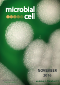
Cover: Colonial growth displayed by the Gram-positive bacteria
Bacillus thuringiensis cultured on sheep blood agar (SBA) medium for 24 hours at 37°C (image by Dr. Todd Parker, Center for Disease Control and Prevention, USA and obtained via the CDC
Public Health Image Library , ID#17110); image modified by MIC. The cover is published under the Creative Commons Attribution (CC BY) license.
Enlarge issue cover
How do yeast sense mitochondrial dysfunction?
Dmitry A. Knorre, Svyatoslav S. Sokolov, Anna N. Zyrina, Fedor F. Severin
Reviews |
page 532-539 | 10.15698/mic2016.11.537 | Full text | PDF |
Abstract
Apart from energy transformation, mitochondria play important signaling roles. In yeast, mitochondrial signaling relies on several molecular cascades. However, it is not clear how a cell detects a particular mitochondrial malfunction. The problem is that there are many possible manifestations of mitochondrial dysfunction. For example, exposure to the specific antibiotics can either decrease (inhibitors of respiratory chain) or increase (inhibitors of ATP-synthase) mitochondrial transmembrane potential. Moreover, even in the absence of the dysfunctions, a cell needs feedback from mitochondria to coordinate mitochondrial biogenesis and/or removal by mitophagy during the division cycle. To cope with the complexity, only a limited set of compounds is monitored by yeast cells to estimate mitochondrial functionality. The known examples of such compounds are ATP, reactive oxygen species, intermediates of amino acids synthesis, short peptides, Fe-S clusters and heme, and also the precursor proteins which fail to be imported by mitochondria. On one hand, the levels of these molecules depend not only on mitochondria. On the other hand, these substances are recognized by the cytosolic sensors which transmit the signals to the nucleus leading to general, as opposed to mitochondria-specific, transcriptional response. Therefore, we argue that both ways of mitochondria-to-nucleus communication in yeast are mostly (if not completely) unspecific, are mediated by the cytosolic signaling machinery and strongly depend on cellular metabolic state.
Sulfur transfer and activation by ubiquitin-like modifier system Uba4•Urm1 link protein urmylation and tRNA thiolation in yeast
André Jüdes, Alexander Bruch, Roland Klassen, Mark Helm, Raffael Schaffrath
Research Articles |
page 554-564 | 10.15698/mic2016.11.539 | Full text | PDF |
Abstract
Urm1 is a unique dual-function member of the ubiquitin protein family and conserved from yeast to man. It acts both as a protein modifier in ubiquitin-like urmylation and as a sulfur donor for tRNA thiolation, which in concert with the Elongator pathway forms 5-methoxy-carbonyl-methyl-2-thio (mcm5s2) modified wobble uridines (U34) in anticodons. Using Saccharomyces cerevisiae as a model to study a relationship between these two functions, we examined whether cultivation temperature and sulfur supply previously implicated in the tRNA thiolation branch of the URM1 pathway also contribute to proper urmylation. Monitoring Urm1 conjugation, we found urmylation of the peroxiredoxin Ahp1 is suppressed either at elevated cultivation temperatures or under sulfur starvation. In line with this, mutants with sulfur transfer defects that are linked to enzymes (Tum1, Uba4) required for Urm1 activation by thiocarboxylation (Urm1-COSH) were found to maintain drastically reduced levels of Ahp1 urmylation and mcm5s2U34 modification. Moreover, as revealed by site specific mutagenesis, the S-transfer rhodanese domain (RHD) in the E1-like activator (Uba4) crucial for Urm1-COSH formation is critical but not essential for protein urmylation and tRNA thiolation. In sum, sulfur supply, transfer and activation chemically link protein urmylation and tRNA thiolation. These are features that distinguish the ubiquitin-like modifier system Uba4•Urm1 from canonical ubiquitin family members and will help elucidate whether, in addition to their mechanistic links, the protein and tRNA modification branches of the URM1 pathway may also relate in function to one another.
The ubiquitin-conjugating enzyme, Ubc1, indirectly regulates SNF1 kinase activity via Forkhead-dependent transcription
Rubin Jiao, Liubov Lobanova, Amanda Waldner, Anthony Fu, Linda Xiao, Troy A. Harkness, and Terra G. Arnason
Research Articles |
page 540-553 | 10.15698/mic2016.11.538 | Full text | PDF |
Abstract
The SNF1 kinase in Saccharomyces cerevisiae is an excellent model to study the regulation and function of the AMP-dependent protein kinase (AMPK) family of serine-threonine protein kinases. Yeast discoveries regarding the regulation of this non-hormonal sensor of metabolic/environmental stress are conserved in higher eukaryotes, including poly-ubiquitination of the α-subunit of yeast (Snf1) and human (AMPKα) that ultimately effects subunit stability and enzyme activity. The ubiquitin-cascade enzymes responsible for targeting Snf1 remain unknown, leading us to screen for those that impact SNF1 kinase function. We identified the E2, Ubc1, as a regulator of SNF1 kinase function. The decreased Snf1 abundance found upon deletion of Ubc1 is not due to increased degradation, but instead is partly due to impaired SNF1 gene expression, arising from diminished abundance of the Forkhead 1/2 proteins, previously shown to contribute to SNF1 transcription. Ultimately, we report that the Fkh1/2 cognate transcription factor, Hcm1, fails to enter the nucleus in the absence of Ubc1. This implies that Ubc1 acts indirectly through transcriptional effects to modulate SNF1 kinase activity.
Threading Granules in Freiburg: 2nd International Symposium on “One Mitochondrion, Many Diseases – Biological and Molecular Perspectives”, a FRIAS Junior Researcher Conference, Freiburg im Breisgau, Germany, March 9th/10th, 2016
Ralf J. Braun, Ralf M. Zerbes, Florian Steinberg, Denis Gris, Verónica I. Dumit
Meeting Reports |
page 565-568 | 10.15698/mic2016.11.540 | Full text | PDF |
Abstract
Altered mitochondrial activities play an important role in many different human disorders, including cancer and neurodegeneration. At the Freiburg Institute of Advanced Studies (FRIAS) Junior Researcher Conference “One Mitochondrion, Many Diseases – Biological and Molecular Perspectives” (University of Freiburg, Freiburg, Germany), junior and experienced researches discussed common and distinct mechanisms of mitochondrial contributions to various human disorders.
Francisella IglG protein and the DUF4280 proteins: PAAR-like proteins in non-canonical Type VI secretion systems?
Claire Lays, Eric Tannier, Thomas Henry
Microreviews |
page 576-578 | 10.15698/mic2016.11.543 | Full text | PDF |
Abstract
Type VI secretion systems (T6SS) are bacterial molecular machines translocating effector proteins into target cells. T6SS are widely present in Gram-negative bacteria where they predominantly act to kill neighboring bacteria. This secretion system is reminiscent of the tail of contractile bacteriophages and consists of a contractile sheath anchored in the bacterial envelope and an inner tube made of stacks of the Hcp protein. The Hcp tube is capped with a VgrG trimer and a spike protein termed PAAR, which acts as the membrane-puncturing device. Francisella tularensis, the agent of tularemia, is an intracellular bacterium replicating within the host cytosol. Upon entry into the host cell, F. tularensis rapidly lyses the host vacuolar membrane to reach the host cytosol. This escape is dependent on the Francisella Pathogenicity Island (FPI), which is encoding an atypical T6SS. Among the 17 proteins encoded by the FPI, most of them required for virulence, eight have some homology to canonical T6SS proteins. We recently identified the function of one protein of unknown function encoded within the FPI, IglG. By three-dimensional modelling and following validation by different techniques, we found that IglG adopts a fold resembling the one of PAAR proteins. Importantly, IglG features a domain of unknown function DUF4280, present in numerous bacterial species. We thus propose to rename this domain of unknown function, PAAR-like domain, and discuss here the characteristics of this domain and its distribution in both Gram-negative and Gram-positive bacteria.
NprR, a moonlighting quorum sensor shifting from a phosphatase activity to a transcriptional activator
Stéphane Perchat, Antoine Talagas, Samira Zouhir, Sandrine Poncet, Laurent Bouillaut, Sylvie Nessler and Didier Lereclus
Microreviews |
page 573-575 | 10.15698/mic2016.11.542 | Full text | PDF |
Abstract
Regulation of biological functions requires factors (proteins, peptides or chemicals) able to sense and translate environmental conditions or any circumstances in order to modulate the transcription of a gene, the stability of a transcript or the activity of a protein. Quorum sensing is a regulation mechanism connecting cell density to the physiological state of a single cell. In bacteria, quorum sensing coordinates virulence, cell fate and commitment to sporulation and other adaptation properties. The critical role of such regulatory systems was demonstrated in pathogenicity and adaptation of bacteria from the Bacillus cereus group (i.e. B. cereus and Bacillus thuringiensis). Furthermore, using insects as a model of infection, it was shown that sequential activation of several quorum sensing systems allowed bacteria to switch from a virulence state to a necrotrophic lifestyle, allowing their survival in the host cadaver, and ultimately to the commitment into sporulation. The chronological development of these physiological states is directed by quorum sensors forming the RNPP family. Among them, NprR combines two distinct functions connecting sporulation to necrotrophism in B. thuringiensis. In the absence of its cognate signaling peptide (NprX), NprR negatively controls sporulation by acting as a phosphatase. In the presence of NprX, it acts as a transcription factor regulating a set of genes involved in the survival of the bacteria in the insect cadaver.
The interaction between herpes simplex virus 1 genome and promyelocytic leukemia nuclear bodies (PML-NBs) as a hallmark of the entry in latency
Patrick Lomonte
Microreviews |
page 569-572 | 10.15698/mic2016.11.541 | Full text | PDF |
Abstract
Herpes simplex virus 1 (HSV-1) is a human pathogen that establishes latency in the nucleus of infected neurons in the PNS and the CNS. At the transcriptional level latency is characterized by a quasi-complete silencing of the extrachromosomal viral genome that otherwise expresses more than 80 genes during the lytic cycle. In neurons, latency is anticipated to be the default transcriptional program; however, limited information exists on the molecular mechanisms that force the virus to enter the latent state. Our recent study demonstrates that the interaction of the viral genomes with the nuclear architecture and specifically the promyelocytic leukemia nuclear bodies (PML-NBs) is a major determinant for the entry of HSV-1 into latency (Maroui MA, Callé A et al. (2016). Latency entry of herpes simplex virus 1 is determined by the interaction of its genome with the nuclear environment. PLoS Pathogens 12(9): e1005834.).


