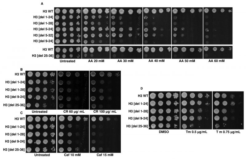
FIGURE 9: Screening of H3 N-terminal deletion mutants identifies acetic acid-hypersensitive mutants. (A) Spot-test assays of the wild-type (H3 WT) and N-terminal tail truncation mutants in different acetic acid concentrations (20, 30, 40, 50, 60 mM). Plates were incubated at 30°C for 72h and photographed. (B) Spot-test assays of the H3 WT and N-terminal tail truncation mutants in different concentrations of Congo red (CR) (60, 100 μg/mL) or (C) Caffeine (Caf) (10, 15 mM). Plates were incubated at 30 °C for 48h (CR) or 72h (Caf) and scanned. (D) Spot-test assays of the H3 WT and N-terminal tail truncation mutants in different concentrations of tunicamycin (Tm) (0.5 μg/mL, 0.75 μg/mL). Images were scanned after 72h.
By continuing to use the site, you agree to the use of cookies. more information
The cookie settings on this website are set to "allow cookies" to give you the best browsing experience possible. If you continue to use this website without changing your cookie settings or you click "Accept" below then you are consenting to this. Please refer to our "privacy statement" and our "terms of use" for further information.

