
Back to article: The extracellular matrix of mycobacterial biofilms: could we shorten the treatment of mycobacterial infections?
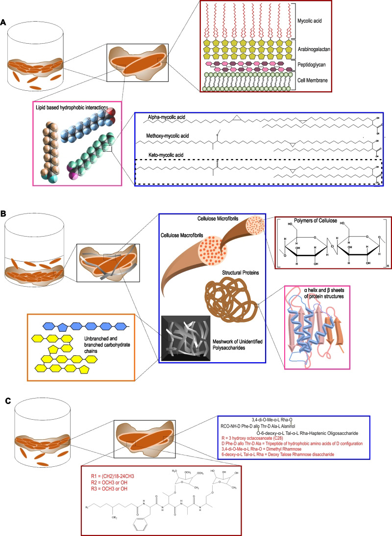
FIGURE 3: Different components of the extracellular polymeric substance of mycobacterial biofilms. (A) The pellicle biofilm model. The inset in maroon shows the different components of the mycobacterial cell wall. The inset in pink shows the lipid based hydrophobic interactions that hold the cells in the biofilm together. The inset in blue shows the different types of mycolic acids present in the extracellular biofilm matrix, the most abundant being keto-mycolic acid. (B) The thiol reductive stress induced submerged biofilm model of Mtb. The inset in blue shows the presence of polysaccharides, cellulose macro- and micro-fibrils, different structural proteins and meshwork of other unidentified branched and unbranched polysaccharides. (C) The pellicle biofilm model of M. smegmatis and M. avium. Apart from mycolic acids, glycopeptidolipids (GPLs) play a major role in pellicle biofilm formation in these bacteria. The inset in maroon depicts the GPL structure in M. smegmatis and the same in blue depicts the GPL structure in M. avium.