
Back to article: Guidelines for DNA recombination and repair studies: Cellular assays of DNA repair pathways
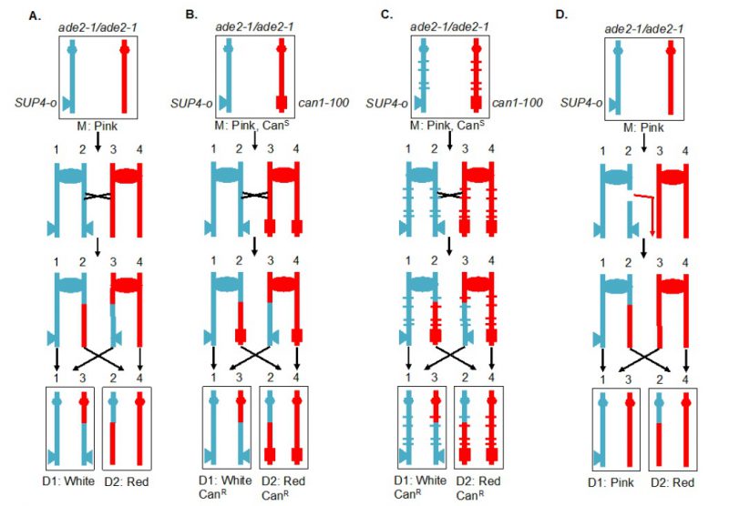
FIGURE 4: Recombination between homologous chromosomes during mitosis. Sectoring assay for monitoring mitotic crossover in the interval between the centromere (oval) and the SUP4-o insertion (triangle). Blue and red indicate the two homologs in the diploid cell. In this version of the assay, the markers are located on the left arm of chromosome V. (A) System for screening mitotic crossover by red/white sectoring assay. (B) Selection system of reciprocal crossovers. (C) Hybrid diploid strains with selection system for recovering reciprocal daughter cells after a crossover event, and with sequence polymorphisms (marked by blue and red ticks) for mapping positions of crossovers. (D) Detection of a non-reciprocal recombination event by a sectoring assay. In the diagram, a DSB on the chromosome containing the SUP4-o gene is repaired by a break-induced replication (BIR) event using the other homolog. The centromere-distal fragment containing the SUP4-o gene is lost, resulting in a pink/red sectored colony.