
Back to article: Depletion of SNAP-23 and Syntaxin 4 alters lipid droplet homeostasis during Chlamydia infection
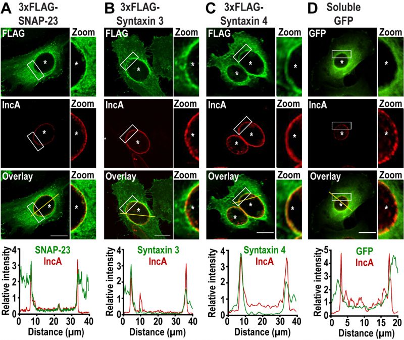
FIGURE 1: SNAP-23, Syntaxin 3, and Syntaxin 4 are recruited to the chlamydial inclusion. HeLa cells were infected with C. trachomatis at a MOI of 2 prior to being transfected with cDNA encoding (A) 3xFLAG-SNAP-23, (B) 3xFLAG-Syntaxin 3, (C) 3xFLAG-Syntaxin 4 or (D) GFP. At 24 h pi, the cells were fixed and labeled with anti-FLAG antibody (green) to label each SNARE and anti-IncA antibody (red) to identify the inclusion membrane. GFP-transfected cells (green) were only labeled with anti-IncA antibody (red). The boxed area of the inclusion membrane within each image is magnified to the right of each image (zoom). Asterisks denote inclusions. Scale bar = 20 μm. Line intensity scans (graphs) were conducted using ImageJ to determine if the FLAG or GFP signal was co-incident with IncA. Yellow line in the overlay image indicates the region of the line scan. Concomitant green (3xFLAG-SNARE or GFP) and red (IncA) peaks signify localization on the inclusion membrane.