Microreviews:
Microbial Cell, Vol. 7, No. 8, pp. 218 - 221; doi: 10.15698/mic2020.08.727
Lipid droplet biogenesis from specialized ER subdomains
1 All India Institute of Medical Sciences (AIIMS), Department of Biotechnology, New Delhi, 110029, India.
2 University of Fribourg, Department of Biology, 1700 Fribourg, Switzerland.
Keywords: ER subdomains, lipid droplet, seipin, Nem1, Yft2, Pex30, diacylglycerol.
Received originally: 01/06/2020 Accepted: 12/06/2020
Published: 16/06/2020
Correspondence:
Vineet Choudhary, All India Institute of Medical Sciences (AIIMS), Department of Biotechnology, New Delhi, 110029, India; vchoudhary@aiims.edu
Roger Schneiter, University of Fribourg, Department of Biology, 1700 Fribourg, Switzerland; roger.schneiter@unifr.ch
Conflict of interest statement: The authors declare that they do not have any compet-ing financial or other interest.
Please cite this article as: Vineet Choudhary and Roger Schneiter (2020). Lipid droplet biogenesis from specialized ER subdomains. Microbial Cell 7(8): 218-221. doi: 10.15698/mic2020.08.727
Lipid droplets (LDs) are cellular compartments dedicated to the storage of metabolic energy in the form of neutral lipids, commonly known as “fat”. The biogenesis of LDs takes place in the endoplasmic reticulum (ER), but its spatial and temporal organization is poorly understood. How exactly sites of LD formation are selected and the succession of proteins and lipids needed to mediate this process remains to be defined. In our current study we show that the yeast triacylglycerol (TAG)-synthases, Lro1 and Dga1 get recruited to discrete ER subdomains where they initiate TAG synthesis and hence LD formation (Choudhary et al. (2020), J Cell Biol). These ER subdomains are defined by yeast seipin, Fld1, and a regulator of diacylglycerol (DAG) production, Nem1. Both Fld1 and Nem1 are ER proteins which localize at contact sites between the ER and LDs. Interestingly, even in cells lacking LDs, Fld1 and Nem1 show punctate localization at ER subdomains independently of each other, but they are required together to recruit the TAG-synthases and hence create functional sites of LD biogenesis. Fld1/Nem1-containing ER subdomains recruit additional LD biogenesis factors, such as Yft2, Pex30, Pet10 and Erg6, and these membrane domains become enriched in DAG. In conclusion, Fld1 and Nem1 play a crucial role in defining ER subdomains for the recruitment of proteins and lipids needed to initiate LD biogenesis.
Lipid droplets (LDs) constitute a widely conserved fat storage compartment that plays a crucial role in cell physiology and metabolism. Growing evidence implicates LDs in many cellular processes, including the endoplasmic reticulum (ER) stress response, protein degradation, membrane trafficking and signal transduction, assembly of infectious viruses, and even for temporary storage of proteins. Defects in LD function in hepatocytes, macrophages and adipocytes leads to pathological conditions, including hepatic steatosis, cardiovascular diseases and obesity. LDs are composed of a core of neutral lipids, mainly triacylglycerols (TAG) and steryl esters (STE), surrounded by a monolayer of phospholipids. The LD surface harbors many lipid metabolic enzymes including lipases and acyltransferases, and structural proteins, such as perilipins (Pet10 in yeast). The biogenesis of LDs takes place in the ER membrane, where the enzymes that catalyze neutral lipid formation are located. The yeast genome encodes two TAG-synthesizing enzymes, Lro1 and Dga1, both convert DAG to TAG and therefore promote LD formation in the ER. TAG and STE are thought to accumulate within the ER bilayer, where they coalesce into lens-like structures. These neutral lipid lenses further grow in size and eventually emerge as LDs towards the cytoplasm, while staying connected to the ER (Fig. 1). Whether these neutral lipid lenses form randomly within the ER or at specific sites, however, remained to be defined.
–
–
Several proteins have been implicated in LD formation, including seipin (Fld1 in yeast), lipin (Pah1 in yeast),
–
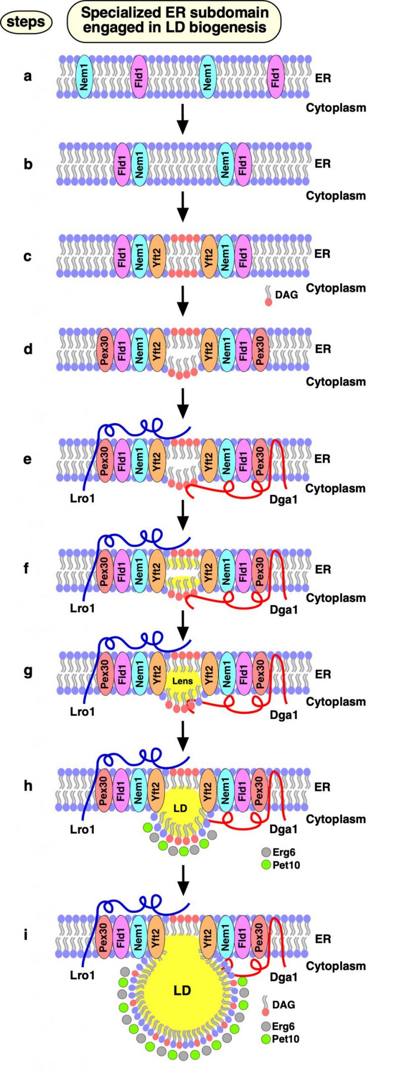
In our recent study, we demonstrate that the biogenesis of LDs occurs at discrete ER subdomains defined by Fld1 and Nem1. Remarkably, localization of Fld1 and Nem1 at these ER subdomains is independent of each other, or of the presence of LDs, but both proteins are required together to create functional sites of LD biogenesis. At these Fld1/Nem1-containing ER subdomains the TAG-synthases, Lro1 or Dga1, get recruited as well as additional factors that promote LD biogenesis, including Yft2, Pex30, and the LD marker proteins Pet10 and Erg6. Proper localization of LD biogenesis in the ER is important, because in cells lacking either Fld1 or Nem1, TAG synthesis occurs ectopically throughout the ER. Ectopically formed LDs in fld1Δ mutant cells do not contain a complete set of LD proteins rendering them functionally impaired. Based on these findings we propose a model for a stepwise initiation of yeast LD biogenesis and an ordered recruitment of proteins to ensure a regulated biogenesis of functional LDs (Fig. 2).
–
–
Apart from the two TAG-synthases, Lro1 and Dga1, yeast expresses two STE synthases, Are1 and Are2. Mutant cells lacking all four of these neutral lipid biosynthetic enzymes (4ΔKO: lro1Δ dga1Δ are1Δ are2Δ) lack the capacity to produce storage lipids and hence have no LDs. All four of these enzymes are localized in the ER, but Dga1 can also localize to the surface of mature LDs. In 4ΔKO cells, Fld1 and Nem1 localize to discrete ER subdomains (Fig. 2, step a). What determines this punctate ER localization of Fld1 and Nem1 has not yet been established, but is likely mediated by protein and/or lipid cues. Most of the Nem1-containing ER puncta appear to be immobile in the ER; some Fld1 puncta show rapid mobility along the ER, while others appear to be stable. The sites where both Fld1 and Nem1 colocalize (Fld1/Nem1 sites) become functional for LD biogenesis (Fig. 2, step b). Induction of TAG synthesis results in an increased colocalization of Fld1 and Nem1, suggesting that the number of LD formation sites is coordinated with the levels of neutral lipids that are being produced. Nem1 activates Pah1 at Fld1/Nem1 sites, which in turn induces an increased local production of DAG (Fig. 2, step c). At this stage, Fld1 might prevent the outward diffusion of DAG into the ER bilayer. Consistent with this notion, DAG appears to be enriched at Fld1/Nem1 sites when cells were stimulated to produce LDs by the addition of oleic acid.
–
At this stage, Yft2 becomes enriched at Fld1/Nem1 sites, possibly due to its direct binding to DAG and/or TAG (Fig. 2, step c). Consistent with this, mammalian FIT2 has previously been shown to bind DAG and TAG in vitro. If Nem1 is missing from Fld1/Nem1 sites, Yft2 fails to get recruited, possibly because of limited DAG and/or TAG levels. In agreement with this hypothesis, overexpression of a hyperactive allele of PAH1 (Pah1-7P), which bypasses the requirement for activation by Nem1, rescues the recruitment of Yft2 at Fld1 sites in cells lacking Nem1. After the association of Yft2 with Fld1/Nem1 sites, the membrane shaping protein Pex30 is recruited (Fig. 2, step d). Pex30 acts downstream of Fld1, Nem1 and Yft2, and its activity at these ER subdomains might promote deformation of the ER bilayer membrane, thereby helping to accommodate even more DAG and/or TAG. Consistent with this notion, previous studies have shown that an ER-anchored DAG sensor is enriched at sites containing Fld1, Nem1, Yft2 and Pex30.
–
Finally, the TAG-synthases, Lro1 or Dga1 get recruited at Fld1/Nem1/Yft2/Pex30 sites, catalyzing the conversion of DAG to TAG and hence formation of neutral lipid lenses and promoting their growth into nascent LDs (Fig. 2, steps e-h). In the absence of either Fld1 or Nem1, these TAG-synthases do not concentrate at discrete spots and instead localize throughout the ER, resulting in ectopic TAG synthesis. Lack of either Fld1 or Nem1 also results in mislocalization of Pex30 and Yft2. Hence, all four of these proteins Fld1/Nem1/Yft2/Pex30 appear to work together at ER subdomains to promote localized synthesis and packaging of neutral lipids. At the very last step, bona fide LD marker proteins, such as the perilipin homolog Pet10 and the ergosterol biosynthetic enzyme Erg6 get recruited to these nascent LDs, thereby stabilizing the LD surface and further promoting their growth and emergence towards the cytoplasm (Fig. 2, steps h, i).
–
In summary, our findings indicate that out of the vast interconnected ER network, ER subdomains defined by the colocalization of Fld1/Nem1/Yft2/Pex30, form sites dedicated to the biogenesis of LDs. Thus, LD formation is a spatially defined process and does not occur through a random coalescence of neutral lipids into lenses in the ER. Importantly, lack of any of the four key players impedes droplet formation and results in aberrant LDs. It will be interesting to see whether similar principles also apply to LD formation in animal cells and whether the human Nem1 orthologue, C-Terminal Domain Nuclear Envelope Phosphatase 1 (CTDNEP1, formerly Dullard), also requires colocalization with seipin to create functional ER subdomains.
ACKNOWLEDGMENTS
We thank all authors who contributed to the original article discussed in this review. This work was supported by the Swiss National Science Foundation (Grant #31003A_17303) and the Novartis Foundation for Medical-Biological Research (grant 19B140). V. Choudhary was supported by European Union’s Horizon 2020 Research and Innovation Programme under the Marie Skłodowska-Curie grant agreement no. 747536.
COPYRIGHT
© 2020

Lipid droplet biogenesis from specialized ER subdomains by Choudhary and Schneiter is licensed under a Creative Commons Attribution 4.0 International License.


