
Back to article: Stable and destabilized GFP reporters to monitor calcineurin activity in Saccharomyces cerevisiae
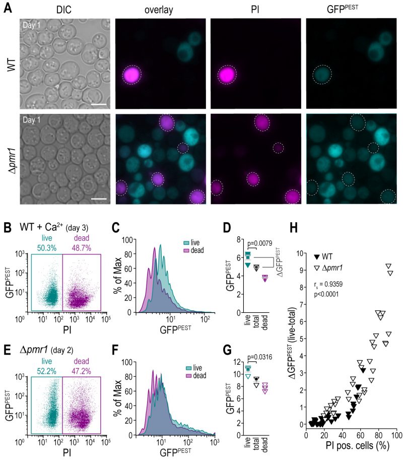
FIGURE 4. A fluorescent reporter for CN activity allows for simultaneous exclusion of dead cells via flow cytometry.(A) Representative micrographs of wild type and Δpmr1 cells equipped with pAMS366-4xCDRE-GFPPEST and stained with propidium iodide (PI) to indicate loss of plasma membrane integrity and thus cell death. Scale bar = 5 µm. (B-G) Flow cytometric analysis of PI-stained aged wild type cells treated with Ca2+ for 1 h as well as Δpmr1 cells equipped with pAMS366-4xCDRE-GFPPEST. Respective dot plots (B, E), histograms of GFPPEST intensities of PI negative (live) and PI positive (dead) cells (C, F) and mean GFPPEST intensities of total, live and dead cell populations (D, G) are shown. (H) Simultaneous quantification of cell death via PI staining and CN activity in cells equipped with pAMS366-4xCDRE-GFPPEST. Wild type and Δpmr1 cells of different age and upon treatment with different concentrations of Ca2+ were analyzed, and the difference of GFPPEST fluorescence intensity between total and live cell populations (ΔGFPPEST (live-total)) was plotted against the percentage of PI positive cells.