
Back to article: Microfluidic techniques for separation of bacterial cells via taxis
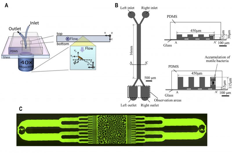
FIGURE 4: Microfluidic device for bacterial rheotaxis. (A) A rectangular channel with a single inlet and outlet was fabricated over an inverted microscope objective lens. Motile bacteria flowed into the channel were tracked using a microscope. Reproduced from [40]. (B) Partition-wall protrusions of height ranging from 30 – 115 µ#x03BC;m were fabricated from PDMS to create the left and right channels. Under controlled flow, motile bacteria flowed into via the left inlet swam towards the right channel and could be collected at the right outlets. Reproduced from [42]. (C) A microfluidic chip was fabricated with microchannels that resembled the texture of porous media. Bacteria were located at the inlets at the extremities of the chip which gradually get distributed into the chip under gravity-driven flow. Reproduced from [43].
40. Kaya T, and Koser H (2012). Direct Upstream Motility in Escherichia coli. Biophys J 102(7): 1514–1523. doi: 10.1016/j.bpj.2012.03.001
42. Ishikawa T, Shioiri T, Numayama-Tsuruta K, Ueno H, Imai Y, and Yamaguchi T (2014). Separation of motile bacteria using drift velocity in a microchannel. Lab Chip 14(5): 1023–1032. doi: 10.1039/C3LC51302E
43. Aufrecht JA, Fowlkes JD, Bible AN, Morrell-Falvey J, Doktycz MJ, and Retterer ST (2019). Pore-scale hydrodynamics influence the spatial evolution of bacterial biofilms in a microfluidic porous network. PLos One 14(6): e0218316. doi: 10.1371/journal.pone.0218316