
Back to article: Cross-species complementation of bacterial- and eukaryotic-type cardiolipin synthases
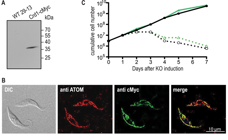
FIGURE 3: Expression, localization, and functionality of ScCrd1 in T. brucei TbCLS conditional knockout cells. (A) SDS-PAGE/immunoblot analysis of whole-cell extracts from wild-type (WT) and Crd1-cMyc-expressing parasites using anti-cMyc antibodies. (B) Immunofluorescence microscopy using antibodies against the endogenous mitochondrial outer membrane protein ATOM (red) and cMyc-tagged ScCrd1 (green). The overlay of both channels (merge) shows overlapping signals (yellow). DIC, Differential Interference Contrast. (C) Growth curve of conditional TbCLS knockout cells expressing ScCrd1. Two different clones (black and green lines) were cultured in the presence (solid lines) or absence (dashed lines) of tetracycline (tet) to maintain or ablate, respectively, TbCLS expression.