
Back to article: Impact of the host on Toxoplasma stage differentiation
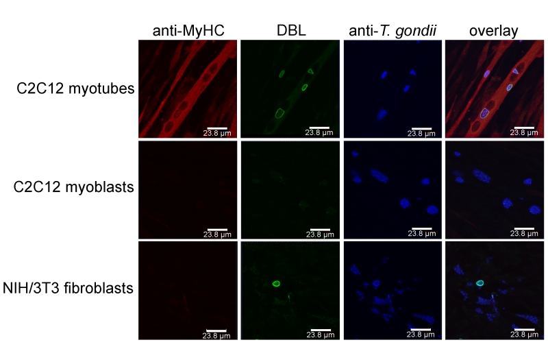
FIGURE 2: T. gondii tissue cyst formation is supported by mature syncytial myotubes but not by proliferating myoblasts or fibroblasts. C2C12 murine skeletal muscle cells (SkMCs) were differentiated in vitro to myotubes. Myotubes, myoblasts and fibroblasts were infected with T. gondii for 72 hours. Efficient myotube formation was verified by staining with an antibody against myosin heavy chain (MyHC), a marker of differentiation to mature SkMCs (red fluorescence). Total parasites irrespective of the parasite stage were labelled with an antiserum (blue fluorescence) and T. gondii tissue cysts were labelled with Dilichos biflorus lectin (DBL) recognizing the tissue cyst wall (green fluorescence). Representative images were recorded by confocal laser scanning microscopy.