
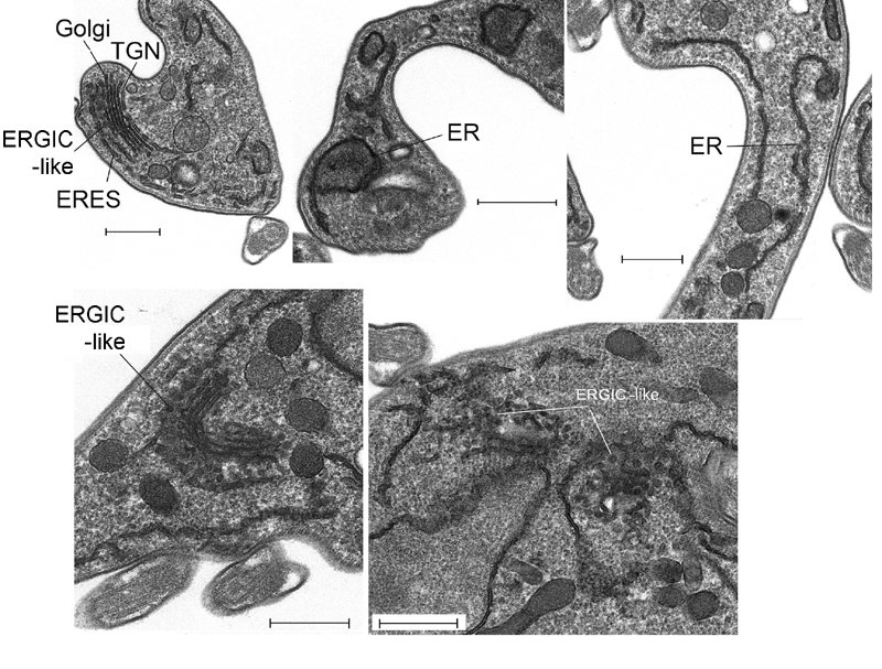
FIGURE 10: Ultrastructural analysis of IGP48 RNAi cells reveals defects in the ER. Transmission electron micrographs of IGP48 RNAi cell lines.
Top left: Representative uninduced IGP48 RNAi cell with major secretory pathway organelles indicated. Other panels are induced cells after 24 hours induction.
Top centre: Distorted ER with apparent lumenal inclusion.
Top right: ER tubules with apparent normal morphology.
Lower left: Extensive vesicles associated with the Golgi complex. Based on observations that the Golgi complex is concave towards the trans-face (see top left panel), these vesicles are likely ER to Golgi transport intermediates corresponding to a structure similar to the ERGIC, i.e. ER-GIC-like.
Lower right: Examples of extensive clusters of vesicles in close association with ER tubules. Scale bars are 500 nm.
Abbreviations: ER, endoplasmic reticulum; ERGIC, ER-Golgi intermediate compartment; TGN, trans-Golgi network.
For figures in higher resolution please refer to PDF version of the article.