
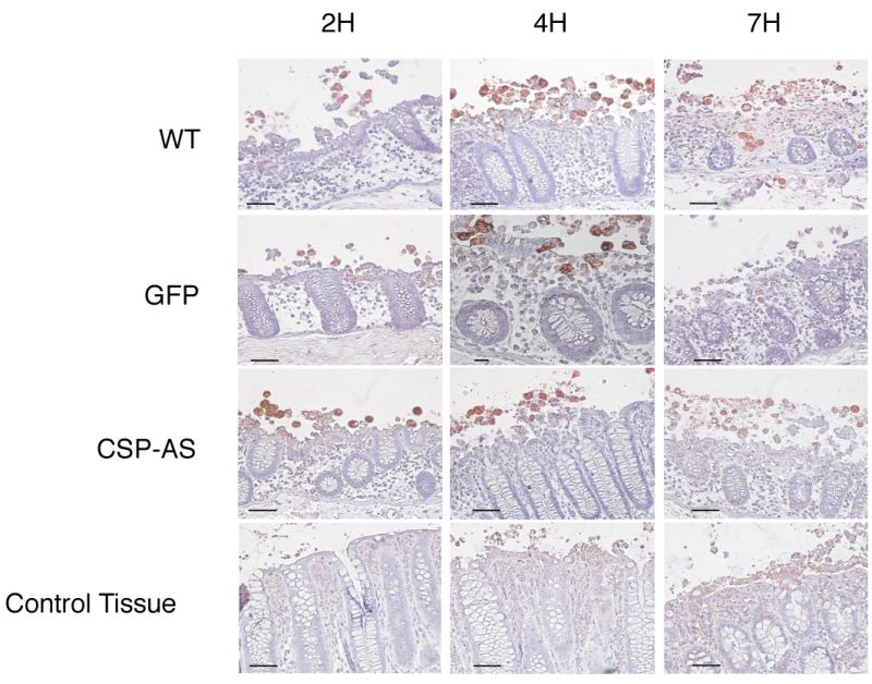
FIGURE 6: Invasion of ex vivo colonic explants was impaired for CSP-AS trophozoites.
Histological examination of colonic tissue sections after 7 h of infection with E. histolytica WT, GFP, CSP-AS strains and in the absence of trophozoites (control). Tissue cross-sections were stained with hematoxylin/eosin reagent. Trophozoites were revealed with an antibody against Gal/GalNAc lectin and appear in red. WT and GFP trophozoites were able to invade the mucosa. In contrast, trophozoites expressing the CSP-AS construct were unable to invade the lamina propria and tended to remain at the surface of the mucosa. Images are representative of three individual experiments. Scale bar: 10 µm.