Table of contents
Volume 9, Issue 2, pp. 24 - 51, February 2022
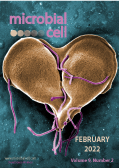
Cover: Digitally-colorized scanning electron microscopic (SEM) image of a Giardia lamblia that was about to become two, separate organisms, as it was caught in a late stage of cell division, producing a heart-shaped form. This protozoan causes the diarrheal disease called giardiasis. Giardia species exist as free-swimming (by means of flagella) trophozoites, and as egg-shaped cysts. It is the cystic stage, which facilitates the survival of these organisms under harsh environmental conditions. The cyst is considered the infective form, and disease is often transmitted by drinking contaminated water (image by Dr. Stan Erlandsen, Center for Disease Control and Prevention, USA and obtained via the CDC Public Health Image Library, ID#11652); image modified by MIC. The cover is published under the Creative Commons Attribution (CC BY) license.
Enlarge issue cover
Endomembrane remodeling and dynamics in Salmonella infection
Ziyan Fang and Stéphane Méresse
Reviews |
page 24-41 | 10.15698/mic2022.02.769 | Full text | PDF |
Abstract
Salmonellae are bacteria that cause moderate to severe infections in humans, depending on the strain and the immune status of the infected host. These pathogens have the particularity of residing in the cells of the infected host. They are usually found in a vacuolar compartment that the bacteria shape with the help of effector proteins. Following invasion of a eukaryotic cell, the bacterial vacuole undergoes maturation characterized by changes in localization, composition and morphology. In particular, membrane tubules stretching over the microtubule cytoskeleton are formed from the bacterial vacuole. Although these tubules do not occur in all infected cells, they are functionally important and promote intracellular replication. This review focuses on the role and significance of membrane compartment remodeling observed in infected cells and the bacterial and host cell pathways involved.
Chromosome-condensed G1 phase yeast cells are tolerant to desiccation stress
Zhaojie Zhang and Gracie R. Zhang
Research Articles |
page 42-51 | 10.15698/mic2022.02.770 | Full text | PDF |
Abstract
The budding yeast Saccharomyces cerevisiae is capable of surviving extreme water loss for a long time. However, less is known about the mechanism of its desiccation tolerance. In this study, we revealed that in an exponential culture, all desiccation tolerant yeast cells were in G1 phase and had condensed chromosomes. These cells share certain features of stationary G0 cells, such as low metabolic level. They were also replicatively young, compared to the desiccation sensitive G1 cells. A similar percentage of chromosome-condensed cells were observed in stationary phase but the condensation level was much higher than that of the log-phase cells. These chromosome-condensed stationary cells were also tolerant to desiccation. However, the majority of the desiccation tolerant cells in stationary phase do not have condensed chromosomes. We speculate that the log-phase cells with condensed chromosome might be a unique feature developed through evolution to survive unpredicted sudden changes of the environment.


