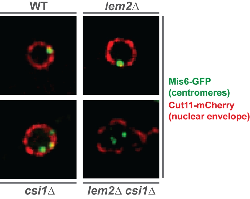
FIGURE 1: Centromeres delocalize from the nuclear periphery in the double mutant lem2∆ csi1∆.
Images show the relative localization of centromeres (green) with respect to the nuclear envelope (red) in WT and indicated mutant nucleus. The kinetochore protein Mis6 is tagged with GFP for visualizing centromeres, while the nuclear pore protein Cut11 is fused to mCherry for detecting the nuclear envelope.

