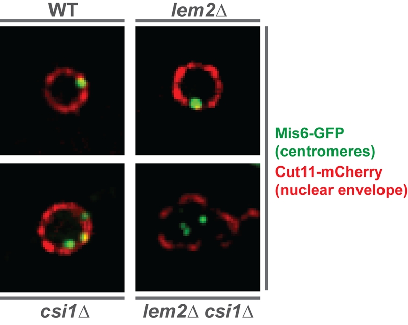
FIGURE 1: Centromeres delocalize from the nuclear periphery in the double mutant lem2csi1∆.

Images show the relative localization of centromeres (green) with respect to the nuclear envelope (red) in WT and indicated mutant nucleus. The kinetochore protein Mis6 is tagged with GFP for visualizing centromeres, while the nuclear pore protein Cut11 is fused to mCherry for detecting the nuclear envelope.

By continuing to use the site, you agree to the use of cookies. more information

The cookie settings on this website are set to "allow cookies" to give you the best browsing experience possible. If you continue to use this website without changing your cookie settings or you click "Accept" below then you are consenting to this. Please refer to our "privacy statement" and our "terms of use" for further information.

Close