
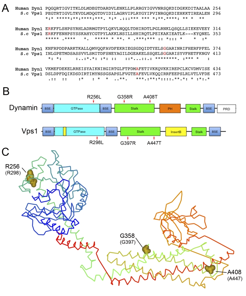
FIGURE 1: Location of Dynamin Mutations. (A) Sequence alignment of relevant region of Dyn-1 and Vps1 showing the position of amino acids selected for mutation. Accession numbers Human Dynamin-1 AAH50279; Vps1 CAA82071. (B) Schematic showing domain structure of mammalian dynamin and yeast Vps1 and corresponding positions of muta-tions. BSE – bundle signaling element, PH – pleckstrin homology, PRD – proline rich domain. (C) The crystal structure of Dynamin-1 (PDB number 3SNH) with dynamin-1 positions denoted with the equivalent mutagenized residues in Vps1 in parentheses.