
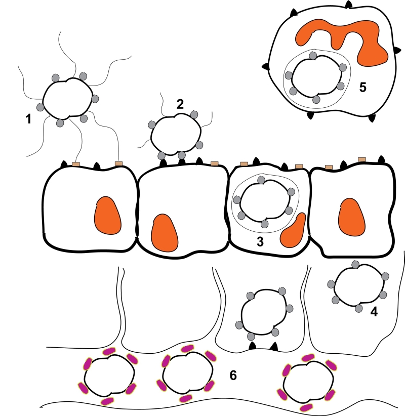
FIGURE 1: Schematic representation of a Neisseria gonorrhoeae infection. 1) Piliated, Opa-expressing gonococci interact with the mucosal epithelium. The thin, hair-like pilus appendages provide the initial contact with receptors on the surface of the mucosal cells. 2) Pili are then retracted which allows for more intimate, Opa-mediated attachment of the bacteria with the CD66 antigens located on the mucosal cells. 3) Following Opa-mediated attachment, the bacteria are engulfed and are internalized into the mucosal cells. 4) Following internalization, some bacteria can transcytose to the basolateral side of the mucosal epithelium. 5) Depending upon which Opa protein is being expressed, gonococci can also reside and survive inside of neutrophils. 6) Following transcytosis, gonococci can enter the bloodstream where heavy sialylation of lipooligosaccharide renders the bacteria serum resistant. This figure is based on [98].
98. Dehio C, Gray-Owen, SD, and Meyer, TF (1998). The role of neisserial Opa proteins in interactions with host cells. Trends Microbiol 6: 489-495. http://dx.doi.org/10.1016/s0966-842x(98)01365-1