
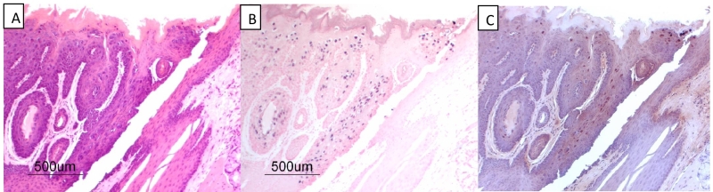
FIGURE 2: MmuPV1 secondary infections of the mouse oral cavity. (A) (H&E), (B) (in situ hybridization using MmuPV1 DNA probe) and (C) (immunohistochemical staining using a monoclonal antibody to MmuPV-1 L1 protein) detecting an infection localized to the base of the tongue at the circumvallate papilla. (A), (B) and (C) are successive 4 μm sections from formalin-fixed paraffin-embedded tissues from athymic mice. One of several examples of MmuPV-1 infection of the circumvallate papilla of the mouse tongue.