
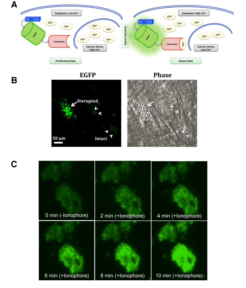
FIGURE 3: GCaMP6s activity in intracellular parasites is induced by natural egress and ionophore. (A) Schematics depicting a rise in cytosolic calcium prior to and/or during egress phase, and detection of calcium by GCaMP6s expressed in the parasite cytosol. The effect of external calcium sources is not illustrated here. (B) GCaMP6s fluorescence in response to the natural egress. Parasitized cells infected with GECI-transgenic parental strain (MOI, 3; 44 h infection) was subjected to EGFP imaging. Image shows intensified EGFP fluorescence during egress (arrow) and background signal in the intact/immature vacuoles (arrowheads). (C) Monitoring of GCaMP6s signal in the parasite cultures treated with ionophore (5 µM A23187). Host cells infected with the parental strain were imaged for 10 min using time-lapse microscopy. Ionophore was added after 1 min of imaging. Still images (snapshots), each with about 2 min intervals, are shown.