
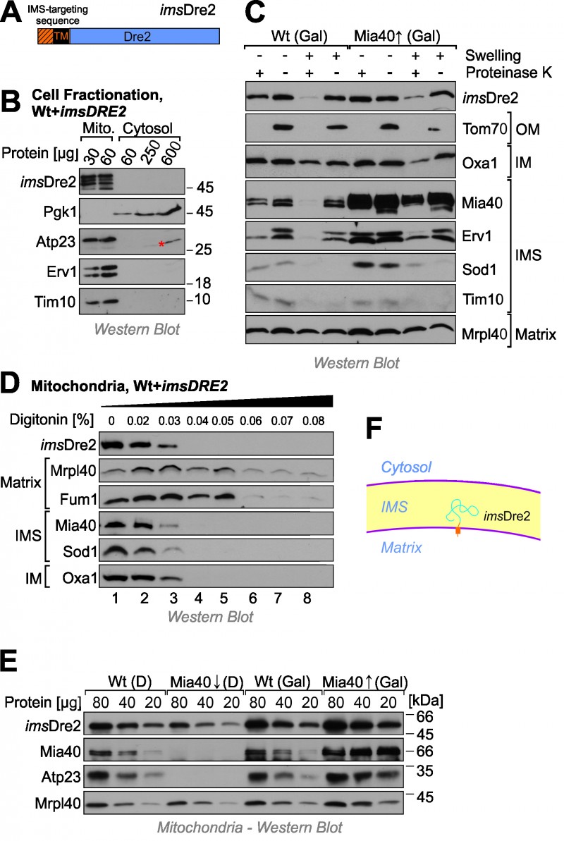
FIGURE 4: Dre2 can be directed into the IMS when fused to an IMS-targeting presequence.
(A) Schematic structure of the imsDre2 fusion protein. TM, transmembrane domain.
(B) Cellular fractionation of imsDre2-overexpressing wild type cells. Some degradation of imsDre2 is visible due to the long exposure time used for this Western blot experiment. The asterisk indicates a crossreaction of the Atp23 antibody.
(C) imsDre2 was expressed in wild type or Mia40-upregulated cells. Mitochondria were isolated and fractionated by hypotonic swelling as described in Fig. 1E.
(D) Digitonin treatment of isolated mitochondria as described in Fig. 1F showing that imsDre2 behaves like an IMS protein.
(E) Steady state levels of imsDre2 and control proteins were analyzed by Western blotting of mitochondria isolated from the indicated strains.
(F) Scheme of the topology of the imsDre2 fusion protein in mitochondria.