
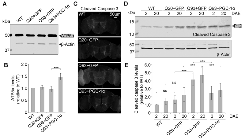
FIGURE 7: DmPGC-1α/spargel suppresses hHttExQ93-induced activation of cell apoptosis.
(A-B) Western blot analysis (A) and quantification (B) of proteins extracted from 2 DAE wild-type, hHttExQ20- or hHttExQ93-overexpressing fly heads. ATP5α was used as a mitochondrial marker. β-Actin was used as an internal control. For quantification, fold change relative to the level of WT group is displayed.
(C) Adult (2 DAE) female brains of wild-type, hHttExQ20- or hHttExQ93-overexpressing flies were probed for cleaved caspase-3. Scale bar is 50 µm.
(D-E) Western blot analysis (D) and quantification (E) of proteins extracted from 2 DAE and 20 DAE wild-type, hHttExQ20- or hHttExQ93-overexpressing fly heads. For quantification, P12 was considered as cleaved caspase-3 (grey box): -fold change relative to each level in 2 DAE wild-type flies. β-Actin was used as an internal control. All data were presented as mean ± S.D., n = 3. Significance level was established by One-Way ANOVA post hoc Bonferroni test. *** P < 0.001.