Table of contents
Volume 5, Issue 4, pp. 169 - 214, April 2018
Cover:
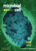
Coxiella burnetii, the bacteria causing the zoonosis Q fever. Shown is a dry fracture electron microscopy image of a
Coxiella burnetii-containing vacuole in a Vero cell. Credit: NIAID; image modified by MIC. The cover is published under the Creative Commons Attribution (CC BY 2.0) license.
Enlarge issue cover
Molecular signature of the imprintosome complex at the mating-type locus in fission yeast
Célia Raimondi, Bernd Jagla, Caroline Proux, Hervé Waxin, Serge Gangloff, Benoit Arcangioli
Research Articles |
page 169-183 | 10.15698/mic2018.04.623 | Full text | PDF |
Abstract
Genetic and molecular studies have indicated that an epigenetic imprint at mat1, the sexual locus of fission yeast, initiates mating type switching. The polar DNA replication of mat1 generates an imprint on the Watson strand. The process by which the imprint is formed and maintained through the cell cycle remains unclear. To understand better the mechanism of imprint formation and stability, we characterized the recruitment of early players of mating type switching at the mat1 region. We found that the switch activating protein 1 (Sap1) is preferentially recruited inside the mat1M allele on a sequence (SS13) that enhances the imprint. The lysine specific demethylases, Lsd1/2, that control the replication fork pause at MPS1 and the formation of the imprint are specifically drafted inside of mat1, regardless of the allele. The CENP-B homolog, Abp1, is highly enriched next to mat1 but it is not required in the process. Additionally, we established the computational signature of the imprint. Using this signature, we show that both sides of the imprinted molecule are bound by Lsd1/2 and Sap1, suggesting a nucleoprotein protective structure defined as imprintosome.
Non-canonical regulation of glutathione and trehalose biosynthesis characterizes non-Saccharomyces wine yeasts with poor performance in active dry yeast production
Esther Gamero-Sandemetrio, Lucía Payá-Tormo, Rocío Gómez-Pastor, Agustín Aranda and Emilia Matallana
Research Articles |
page 184-197 | 10.15698/mic2018.04.624 | Full text | PDF |
Abstract
Several yeast species, belonging to Saccharomyces and non-Saccharomyces genera, play fundamental roles during spontaneous must grape fermentation, and recent studies have shown that mixed fermentations, co-inoculated with S. cerevisiae and non-Saccharomyces strains, can improve wine organoleptic properties. During active dry yeast (ADY) production, antioxidant systems play an essential role in yeast survival and vitality as both biomass propagation and dehydration cause cellular oxidative stress and negatively affect technological performance. Mechanisms for adaptation and resistance to desiccation have been described for S. cerevisiae, but no data are available on the physiology and oxidative stress response of non-Saccharomyces wine yeasts and their potential impact on ADY production. In this study we analyzed the oxidative stress response in several non-Saccharomyces yeast species by measuring the activity of reactive oxygen species (ROS) scavenging enzymes, e.g., catalase and glutathione reductase, accumulation of protective metabolites, e.g., trehalose and reduced glutathione (GSH), and lipid and protein oxidation levels. Our data suggest that non-canonical regulation of glutathione and trehalose biosynthesis could cause poor fermentative performance after ADY production, as it corroborates the corrective effect of antioxidant treatments, during biomass propagation, with both pure chemicals and food-grade argan oil.
Impact of F1Fo-ATP-synthase dimer assembly factors on mitochondrial function and organismic aging
Nadia G Rampello, Maria Stenger, Benedikt Westermann, Heinz D Osiewacz
Research Reports |
page 198-207 | 10.15698/mic2018.04.625 | Full text | PDF |
Abstract
In aerobic organisms, mitochondrial F1Fo-ATP-synthase is the major site of ATP production. Beside this fundamental role, the protein complex is involved in shaping and maintenance of cristae. Previous electron microscopic studies identified the dissociation of F1Fo-ATP-synthase dimers and oligomers during organismic aging correlating with a massive remodeling of the mitochondrial inner membrane. Here we report results aimed to experimentally proof this impact and to obtain further insights into the control of these processes. We focused on the role of the two dimer assembly factors PaATPE and PaATPG of the aging model Podospora anserina. Ablation of either protein strongly affects mitochondrial function and leads to an accumulation of senescence markers demonstrating that the inhibition of dimer formation negatively influences vital functions and accelerates organismic aging. Our data validate a model that links mitochondrial membrane remodeling to aging and identify specific molecular components triggering this process.
Helicobacter hepaticus polysaccharide induces an anti-inflammatory response in intestinal macrophages
Camille Danne and Fiona Powrie
Microreviews |
page 208-211 | 10.15698/mic2018.04.626 | Full text | PDF |
Abstract
A high density of microbes inhabits the intestine, helping with food digestion, vitamin synthesis, xenobiotic detoxification, pathogen resistance and immune system maturation. Crucial for human health, communities of commensal bacteria (collectively termed microbiota) benefit in return from a nutrient-rich environment. Host-microbiota mutualism results from a long-term co-adaptation. At barrier surfaces, immune cells distinguish harmful from commensal bacteria and tolerate non-self organisms at close proximity to the mucosa; gut inhabitants have developed strategies to ensure beneficial conditions in their preferred niche. So far, the complex dialogue of host-microbial mutualism is poorly understood. Helicobacter hepaticus is a member of the mouse microbiota that colonizes the lower intestine without inducing immune pathology. However, when there is a host maladaptation such as the absence of the anti-inflammatory cytokine interleukin 10 (IL-10) or its receptor IL-10R, H. hepaticus triggers aberrant IL-23-driven intestinal inflammation. This response results in major changes in the intestinal innate cell compartment, with the accumulation of inflammatory macrophages. Relying both on a bacterial trigger and on an immune defect, H. hepaticus-induced colitis in the context of IL-10/IL-10R axis deficiency shares many features of human inflammatory bowel diseases (IBD). In our study [Danne et al, Cell Host Microbe 22(6):733-745], we questioned the interactions between H. hepaticus and intestinal macrophages that promote mutualism. Our results show that H. hepaticus produces a large polysaccharide that triggers IL-10 production without a corresponding inflammatory response in macrophages. Moreover, H. hepaticus polysaccharide specifically induces an anti-inflammatory gene signature in vitro and in vivo, including transcriptional factors known as repressors of immune activation. This anti-inflammatory program depends on the TLR2/MSK/CREB pathway, which might be crucial to maintain mutualistic relationships at the intestinal interface.
Endolysosomal pathway activity protects cells from neurotoxic TDP-43
Christine Leibiger, Jana Deisel, Andreas Aufschnaiter, Stefanie Ambros, Maria Tereshchenko, Bert M. Verheijen, Sabrina Büttner, and Ralf J. Braun
Microreviews |
page 212-214 | 10.15698/mic2018.04.627 | Full text | PDF |
Abstract
The accumulation of protein aggregates in neurons is a typical pathological hallmark of the motor neuron disease amyotrophic lateral sclerosis (ALS) and of frontotemporal dementia (FTD). In many cases, these aggregates are composed of the 43 kDa TAR DNA-binding protein (TDP‑43). Using a yeast model for TDP‑43 proteinopathies, we observed that the vacuole (the yeast equivalent of lysosomes) markedly contributed to the degradation of TDP‑43. This clearance occurred via TDP‑43-containing vesicles fusing with the vacuole through the concerted action of the endosomal-vacuolar (or endolysosomal) pathway and autophagy. In line with its dominant role in the clearance of TDP‑43, endosomal-vacuolar pathway activity protected cells from the detrimental effects of TDP‑43. In contrast, enhanced autophagy contributed to TDP‑43 cytotoxicity, despite being involved in TDP‑43 degradation. TDP‑43’s interference with endosomal-vacuolar pathway activity may have two deleterious consequences. First, it interferes with its own degradation via this pathway, resulting in TDP‑43 accumulation. Second, it affects vacuolar proteolytic activity, which requires endosomal-vacuolar trafficking. We speculate that the latter contributes to aberrant autophagy. In sum, we propose that ameliorating endolysosomal pathway activity enhances cell survival in TDP‑43-associated diseases.


