Table of contents
Volume 3, Issue 1, pp. 1 - 52, January 2016
Cover:
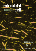
Pseudomonas aeruginosa, an opportunistic bacterial pathogen, can produce large amounts of filamentous bacteriophage during growth as a biofilm. Filamentous bacteriophage produced by
P. aeruginosa interact with host and microbial polymers to assemble liquid crystals. When the biofilm matrix is organized into a liquid crystalline structure, desiccation survival and antibiotic tolerance are promoted. Pictured, fluorescently labeled filamentous bacteriophage (yellow) spontaneously assemble liquid crystalline networks in the presence of the host polymer hyaluronan. Image by Patrick Secor (University of Washington, USA); modified by MIC. The cover is published under the Creative Commons Attribution (CC BY) license.
Enlarge issue cover
Histone modifications as regulators of life and death in Saccharomyces cerevisiae
Birthe Fahrenkrog
Reviews |
page 1-13 | 10.15698/mic2016.01.472 | Full text | PDF |
Abstract
Apoptosis or programmed cell death is an integrated, genetically controlled suicide program that not only regulates tissue homeostasis of multicellular organisms, but also the fate of damaged and aged cells of lower eukaryotes, such as the yeast Saccharomyces cerevisiae. Recent years have revealed key apoptosis regulatory proteins in yeast that play similar roles in mammalian cells. Apoptosis is a process largely defined by characteristic structural rearrangements in the dying cell that include chromatin condensation and DNA fragmentation. The mechanism by which chromosomes restructure during apoptosis is still poorly understood, but it is becoming increasingly clear that altered epigenetic histone modifications are fundamental parameters that influence the chromatin state and the nuclear rearrangements within apoptotic cells. The present review will highlight recent work on the epigenetic regulation of programmed cell death in budding yeast.
Ergosterone-coupled Triazol molecules trigger mitochondrial dysfunction, oxidative stress, and acidocalcisomal Ca2+ release in Leishmania mexicana promastigotes
Figarella K, Marsiccobetre S, Arocha I, Colina W, Hasegawa M, Rodriguez M, Rodriguez-Acosta A, Duszenko M, Benaim G, Uzcategui NL
Research Articles |
page 14-28 | 10.15698/mic2016.01.471 | Full text | PDF |
Abstract
The protozoan parasite Leishmania causes a variety of sicknesses with different clinical manifestations known as leishmaniasis. The chemotherapy currently in use is not adequate because of their side effects, resistance occurrence, and recurrences. Investigations looking for new targets or new active molecules focus mainly on the disruption of parasite specific pathways. In this sense, ergosterol biosynthesis is one of the most attractive because it does not occur in mammals. Here, we report the synthesis of ergosterone coupled molecules and the characterization of their biological activity on Leishmania mexicana promastigotes. Molecule synthesis involved three steps: ergosterone formation using Jones oxidation, synthesis of Girard reagents, and coupling reaction. All compounds were obtained in good yield and high purity. Results show that ergosterone-triazol molecules (Erg-GTr and Erg-GTr2) exhibit an antiproliferative effect in low micromolar range with a selectivity index ~10 when compared to human dermic fibroblasts. Addition of Erg-GTr or Erg-GTr2 to parasites led to a rapid [Ca2+]cyt increase and acidocalcisomes alkalinization, indicating that Ca2+ was released from this organelle. Evaluation of cell death markers revealed some apoptosis-like indicators, as phosphatidylserine exposure, DNA damage, and cytosolic vacuolization and autophagy exacerbation. Furthermore, mitochondrion hyperpolarization and superoxide production increase were detected already 6 hours after drug addition, denoting that oxidative stress is implicated in triggering the observed phenotype. Taken together our results indicate that ergosterone-triazol coupled molecules induce a regulated cell death process in the parasite and may represent starting point molecules in the search of new chemotherapeutic agents to combat leishmaniasis.
Global translational impacts of the loss of the tRNA modification t6A in yeast
Patrick C. Thiaville, Rachel Legendre, Diego Rojas-Benítez, Agnès Baudin-Baillieu, Isabelle Hatin, Guilhem Chalancon, Alvaro Glavic, Olivier Namy, Valérie de Crécy-Lagard
Research Articles |
page 29-45 | 10.15698/mic2016.01.473 | Full text | PDF |
Abstract
The universal tRNA modification t6A is found at position 37 of nearly all tRNAs decoding ANN codons. The absence of t6A37 leads to severe growth defects in baker’s yeast, phenotypes similar to those caused by defects in mcm5s2U34 synthesis. Mutants in mcm5s2U34 can be suppressed by overexpression of tRNALysUUU, but we show t6A phenotypes could not be suppressed by expressing any individual ANN decoding tRNA, and t6A and mcm5s2U are not determinants for each other’s formation. Our results suggest that t6A deficiency, like mcm5s2U deficiency, leads to protein folding defects, and show that the absence of t6A led to stress sensitivities (heat, ethanol, salt) and sensitivity to TOR pathway inhibitors. Additionally, L-homoserine suppressed the slow growth phenotype seen in t6A-deficient strains, and proteins aggregates and Advanced Glycation End-products (AGEs) were increased in the mutants. The global consequences on translation caused by t6A absence were examined by ribosome profiling. Interestingly, the absence of t6A did not lead to global translation defects, but did increase translation initiation at upstream non-AUG codons and increased frame-shifting in specific genes. Analysis of codon occupancy rates suggests that one of the major roles of t6A is to homogenize the process of elongation by slowing the elongation rate at codons decoded by high abundance tRNAs and I34:C3 pairs while increasing the elongation rate of rare tRNAs and G34:U3 pairs. This work reveals that the consequences of t6A absence are complex and multilayered and has set the stage to elucidate the molecular basis of the observed phenotypes.
Biofilm assembly becomes crystal clear – filamentous bacteriophage organize the Pseudomonas aeruginosa biofilm matrix into a liquid crystal
Patrick R. Secor, Laura K. Jennings, Lia A. Michaels, Johanna M. Sweere, Pradeep K. Singh, William C. Parks, Paul L. Bollyky
Microreviews |
page 49-52 | 10.15698/mic2016.01.475 | Full text | PDF |
Abstract
Pseudomonas aeruginosa is an opportunistic bacterial pathogen associated with many types of chronic infection. At sites of chronic infection, such as the airways of people with cystic fibrosis (CF), P. aeruginosa forms biofilm-like aggregates. These are clusters of bacterial cells encased in a polymer-rich matrix that shields bacteria from environmental stresses and antibiotic treatment. When P. aeruginosa forms a biofilm, large amounts of filamentous Pf bacteriophage (phage) are produced. Unlike most phage that typically lyse and kill their bacterial hosts, filamentous phage of the genus Inovirus, which includes Pf phage, often do not, and instead are continuously extruded from the bacteria. Here, we discuss the implications of the accumulation of filamentous Pf phage in the biofilm matrix, where they interact with matrix polymers to organize the biofilm into a highly ordered liquid crystal. This structural configuration promotes bacterial adhesion, desiccation survival, and antibiotic tolerance – all features typically associated with biofilms. We propose that Pf phage make structural contributions to P. aeruginosa biofilms and that this constitutes a novel form of symbiosis between bacteria and bacteriophage.
Spermidine cures yeast of prions
Shaun H. Speldewinde, Chris M. Grant
Microreviews |
page 46-48 | 10.15698/mic2016.01.474 | Full text | PDF |
Abstract
Prions are self-perpetuating amyloid protein aggregates which underlie various neurodegenerative diseases in mammals. The molecular basis underlying their conversion from a normally soluble protein into the prion form remains largely unknown. Studies aimed at uncovering these mechanism(s) are therefore essential if we are to develop effective therapeutic strategies to counteract these disease-causing entities. Autophagy is a cellular degradation system which has predominantly been considered as a non-selective bulk degradation process which recycles macromolecules in response to starvation conditions. We now know that autophagy also serves as a protein quality control mechanism which selectively degrades protein aggregates and damaged organelles. These are commonly accumulated in various neurodegenerative disorders including prion diseases. In our recent study [Speldewinde et al. Mol. Biol. Cell. (2015)] we used the well-established yeast [PSI+]/Sup35 and [PIN+]/Rnq1 prion models to show that autophagy prevents sporadic prion formation. Importantly, we found that spermidine, a polyamine that has been used to increase autophagic flux, acts as a protective agent which prevents spontaneous prion formation.


